

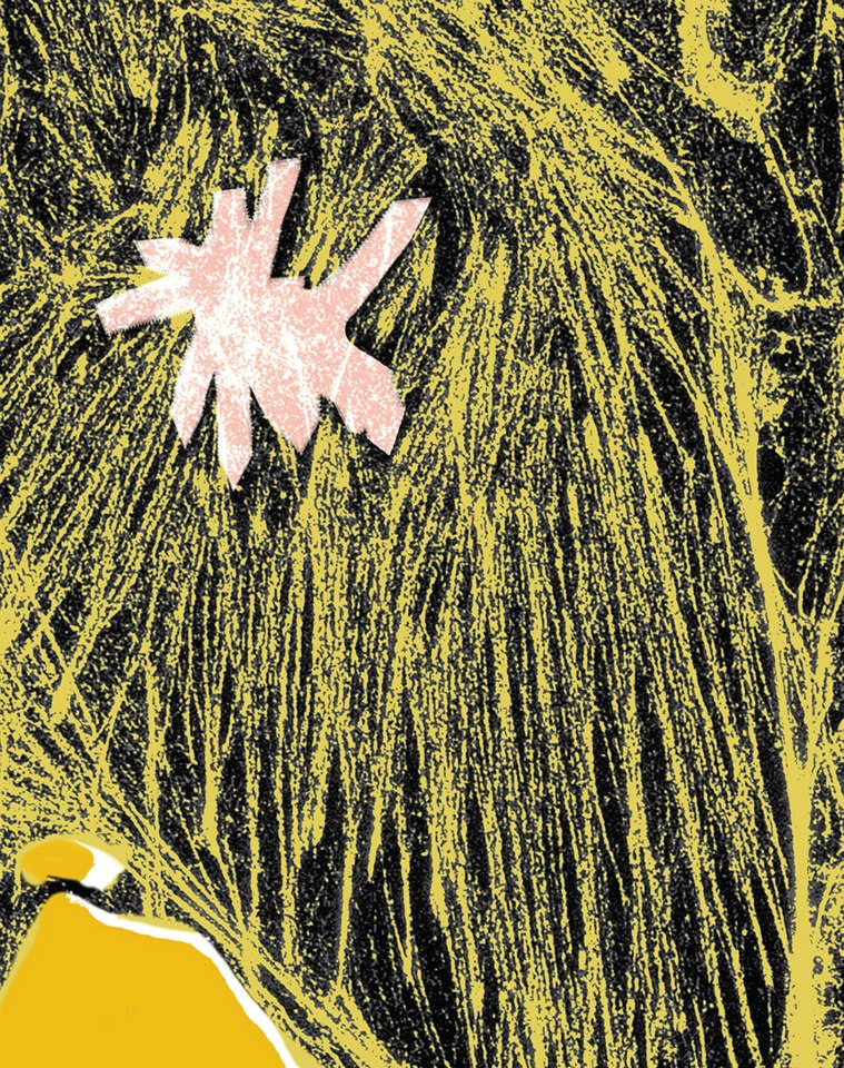
Zeiss Elyra Structured Illumination Superresolution System
Image provided by Mayandi Sivaguru, Core Facilities
Research funded by the Institute for Genomic Biology
This image of the internal structure, or cytoskeleton, of a cell was produced through super-resolution microscopy. This new technique defies the physical limits of conventional microscopy and reveals twice the detail that can be obtained with older techniques. The higher level of detail helps researchers understand the structure and mechanics of much smaller objects.