

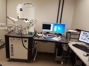
The SBF-SEM is an automated technique for obtaining serial block face images and 3D data using a SEM. Our Zeiss Sigma VP 3View system is a Gatan 3View SBF microtome housed in a Gemini SEM column. The microtome is equipped with a diamond knife which is mounted inside the chamber of the SEM and shaves of <50nm (15 – 200nm) of the sample in between imaging. Images are collected using a backscattered (BD) electron detector which results in a TEM-like image. The SBF-SEM imaging process is fully automated, allowing for a large volume acquisition without the risk of losing sections.
The instrument has the following features and advantages:
-
This Zeiss Sigma VP 3View system is a Gatan 3View SBF microtome housed in a Gemini SEM column.
-
It is equipped with several detectors for both regular SEM imaging and SBF imaging. It includes an in-lens detector for detecting secondary (SE) and/or backscattered (BSE) electrons as well as a Variable Pressure Secondary Electron (VPSE G3) detector.
-
The SMART SEM software is for imaging in SEM mode and the Gatan Microscopy Suite (also known as Gatan Digital Micrograph) is for SBF imaging and data analysis.
-
<50nm X, Y stage repeatability allows multi-region imaging without losing data due to imprecise stage motion.
-
You can automatically acquire images across multiple regions of the sample utilizing multi-regions of interest (mROIs).
-
Large format image support (up to 32k x 24k) allows very large images to be collected.
-
15nm Z section thickness without the need to unblur multi-kV images
-
High performance back-scatter detector allows high speed imaging at low kV without giving up image quality
Links:
-
Zeiss Sigma 3View Introduction Video: https://www.youtube.com/watch?v=3yk3rbgu49o
-
Gatan 3View Technical Notes and Protocols: http://www.gatan.com/products/sem-imaging-spectroscopy/3view-system
-
Zeiss Sigma 3View White Paper: http://applications.zeiss.com/C125792900358A3F/0/12129D5013398101C1257A91004312DF/
-
Zeiss Sigma 3View Webinar: https://www.zeiss.com/microscopy/us/products/scanning-electron-microscopes/merlin-life-science.html
-
3View Sample Processing Request Form:
Manufacturer: Zeiss
Equipment Model: Sigma VP
Location: Room 15, Carl R. Woese Institute for Genomic Biology
Contact Person: Kingsley Boateng, 217-333-1642, kboateng@igb.illinois.edu
Fees & Consumables: See fees page
Sample Images and Videos:
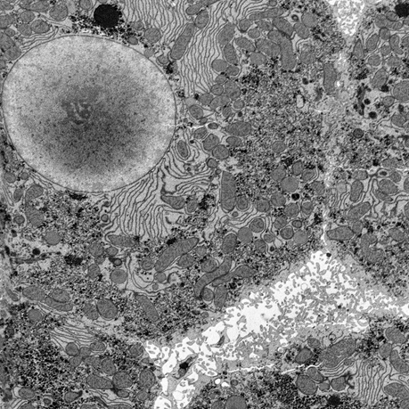
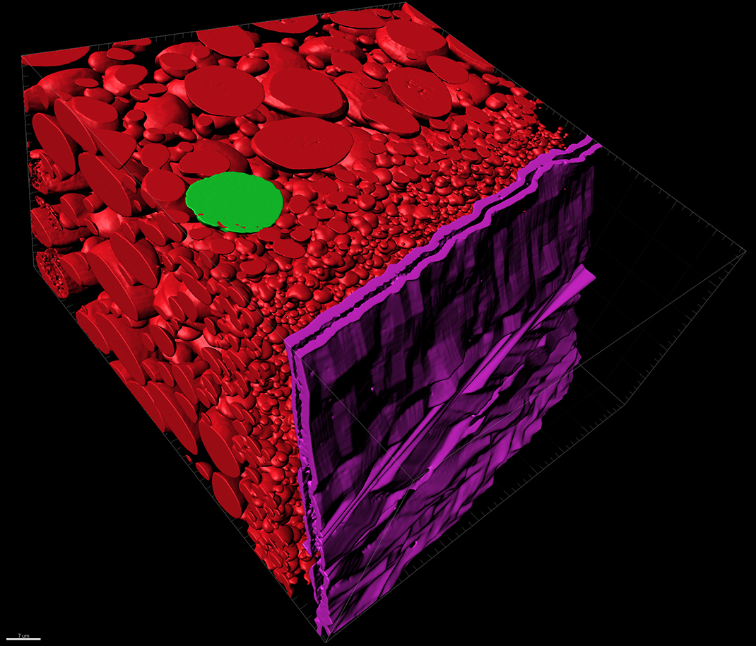
Liver Tissue
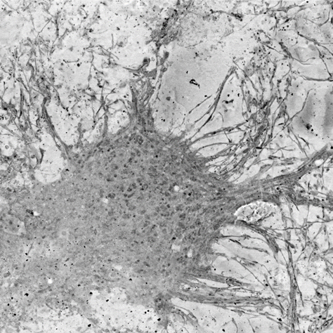
Neurons
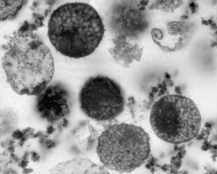
Archaea cells
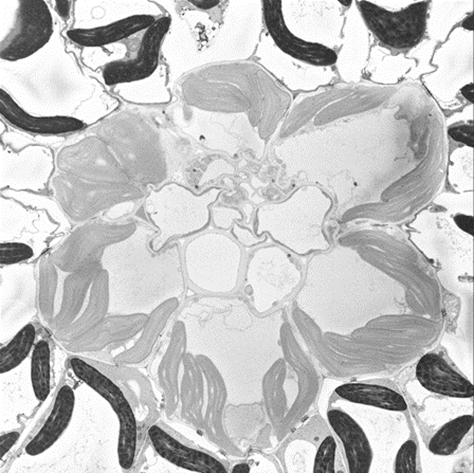
Plant leaf - chloroplast
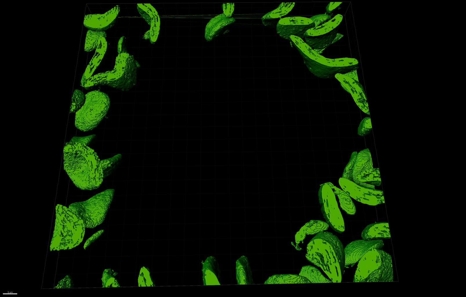
Segmented chloroplast with MIB Software
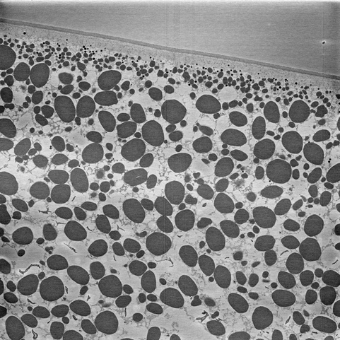
Xenopus laevis oocytes (frog egg yoke)
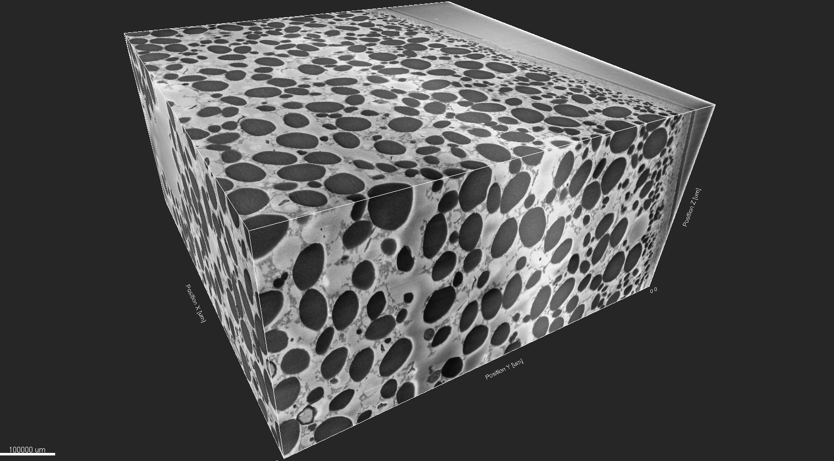
Segmented frog egg-yoke with MIB Software