Kingsley Boateng, Stephen Fleming, and Ryan Dilger
Ryan Dilger Laboratory
Funded by IGB
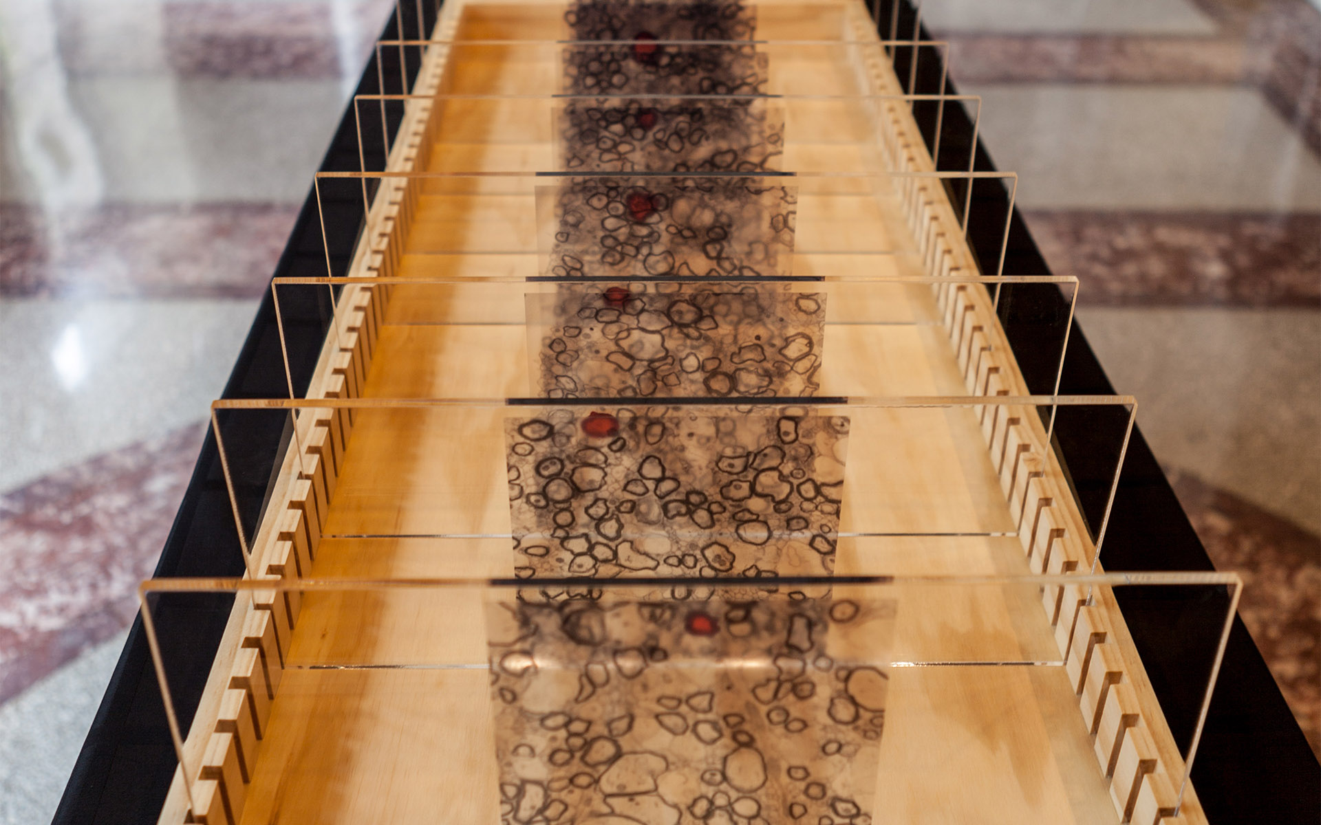
In an effort to understand the complex processes of the brain, researchers are leveraging novel imaging technologies to create increasingly detailed models of brain structure. The images seen in this piece were created when researchers repeatedly imaged the surface of a sample of brain tissue from a young pig, then removed a thin layer of tissue before imaging again. The extended projections of neurons, called axons, appear as dark rings. One axon is highlighted in red.
This serial imaging produces images that enable the viewer to construct an idea of the true shape of the tissue. Each slice that is removed adds a new layer of information to the growing model; thus two dimensional objects grow into structures that can be traced through three dimensional space.

