| Posters | ||
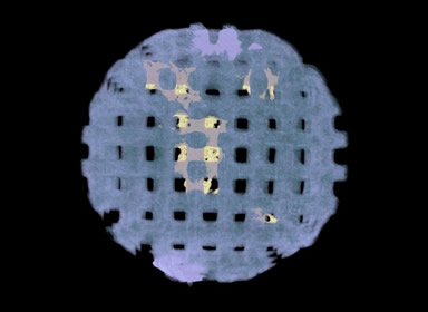
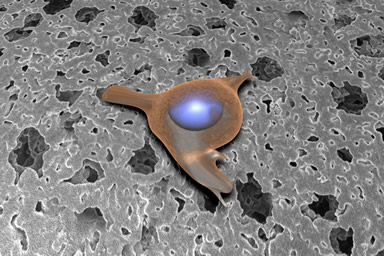
Bone Scaffold Implants The above image is a complete hydroxyapetite bone scaffold after eight weeks as an implant in living tissue. The light purple coloring is the scaffold itself, while the yellow is bone that has grown in the scaffold. The scaffold was imaged using the SkyScan 1172 micro-CT system, and the image was created and analyzed using Amira, both in the VMIL. |
||
Cell Attachment to Implant Surface This image depicts one frame from an animation that illustrates the development of an artificial bone implant. The animation, which was produced using Maya, Analyze, Final Cut Pro, and other tools in the VMIL, walks the viewer through the Mandible Reconstruction Project; an ongoing, multi-disciplinary effort to develop an alternative approach to bone replacement. In this image, bone cells migrate across the surface of the implant, which by design has a microporosity meant to encourage this action. |
||
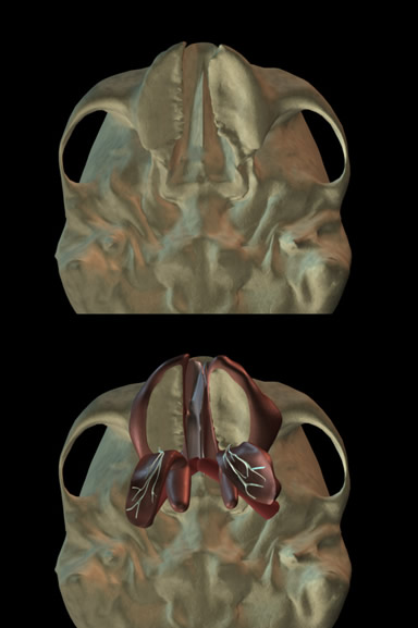
Computer Modeling of the Levator Veli Palatini Muscle A child born with a cleft palate may undergo multiple surgeries to restore anatomy and regain muscle function. In numerous cases, even after surgical intervention to repair the cleft palate, normal sounding speech may not be restored due to improper surgical alignment of a palatal muscle known as the levator veli palatini (levator). Success in surgery is contingent upon a better understanding of the structures and functions of the levator muscle and surrounding soft tissue. The goal of the project is to demonstrate a proof of concept in the use of MRI and 3D computer technology for the purposes of presurgical planning, assessment of levator muscle morphology before and after surgery, and postsurgical assessments of the levator muscle. In this instance, the image represents a view of the undersurface of the skull with the mandible removed, and demonstrates the cleft of the palate and the surgical procedure used to reconstruct the internal anatomy. This research is a step closer to providing significant advances in the clinical realm such as reducing human error during surgery, computer simulation of surgical planning, as well as patient specific modeling, diagnosing, and treatment for individuals born with a cleft palate. This project demonstrates the implementation of translational research by combining basic research with applied clinical research. |
||
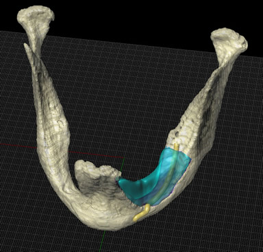
Mandibular Reconstruction The blue region of this image depicts an area of significant bone loss in a human jaw that has occurred as a result of gradual reabsorption of the bone material over time. Loss of teeth, disease, or other trauma may result in the loss of bone if not carefully monitored. In this instance, degradation of the jaw has exposed the nerve (in yellow) resulting in severe discomfort for the patient. The reconstruction of missing mandibular material was created by Janet Sinn-Hanlon from original CT data in the software packages Analyze and Rhino, available in the VMIL. Rhino created the rendering shown here.
|
||
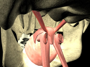
3D Computer Reconstruction of the Velopharyngeal MechanismThe goal of this project was to create a 3D model of velopharyngeal mechanism based on MRI data to demonstrate the velum at rest and during elevation. Using quantitative data, length, width, origin and insertion of the major muscles of the velopharynx were modeled using the Maya Unlimited 6.5 software system. The model was animated to demonstrate velar movement during repeated sustained vowel production. Improvements in visualization of the velopharynx through 3D computer graphics offer a promising future for the field of speech and hearing science. |
||
|
Deposition of scaffold
|
||
| home | research | publications | CV | links |