Novel imaging techniques are necessary for examining whole brain protein expression patterns. Animal brains are large, complex structures that are difficult to image comprehensively on thin sections by traditional immunohistochemistry (IHC) techniques.
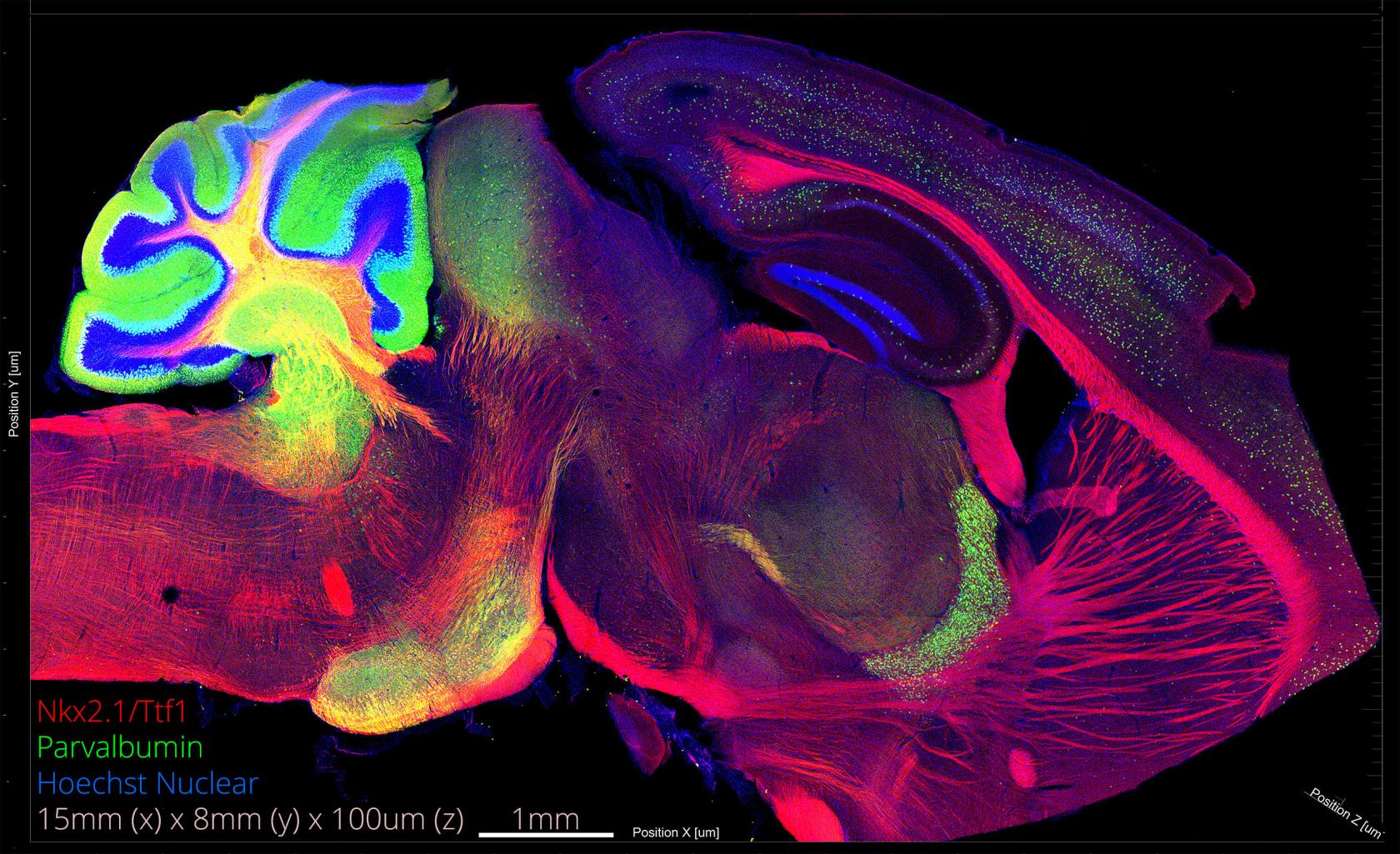
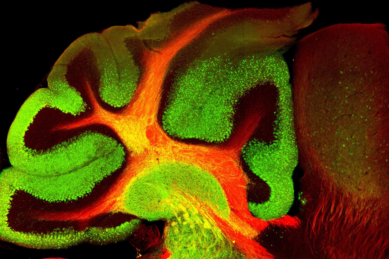
This image shows a 100um thick mouse brain slice, cleared with optimized CLARITY techniques for imaging on the IGB LSM 710 confocal microscope at 10x in three channels, with a tiling size of 15x9 and a z-stack of 10. The image was then rendered in 3D with Imaris x8.
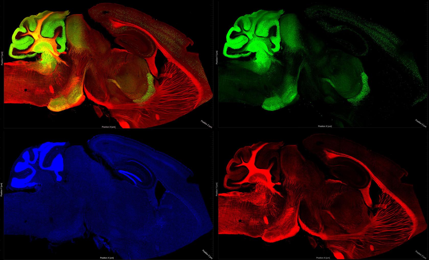
The blue channel indicates nuclei with Hoechst stain. The green channel identifies GABAergic inhibitory interneurons with IHC for parvalbumin.
The red channel shows IHC for a transcription factor involved in neuron differentiation and social learning, Ttf1.
These exciting techniques allow imaging of complete brain structures while still allowing for cellular level resolution.