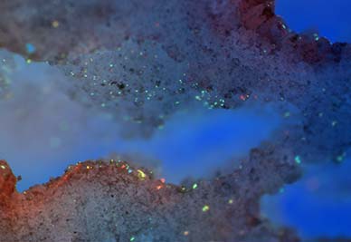
  • February 2013

    Fast Fourier Transformation of a Superresolution Structured Illumination Image of a Cell

    Structured illumination superresolution imaging technique breaks the diffraction limit of light by half in optical microscopy which was held over a century. Cellular structures and proteins now could be visualized up to 120 nm in XY and 250 nm in Z axis. By creating Moiré fringes using a known patterned illumination over the samples information beyond the conventional field of view is transferred into the actual viewable field. This enhanced information is calculated in the frequency space using a set of sophisticated Fast Fourier Transformation (FFT) algorithms. The image is a three dimensional FFT of 19 optical sections of a cell labeled for visualizing actin in single fluorescent channel processed to emphasize the signal-to-noise ratio in 3D.