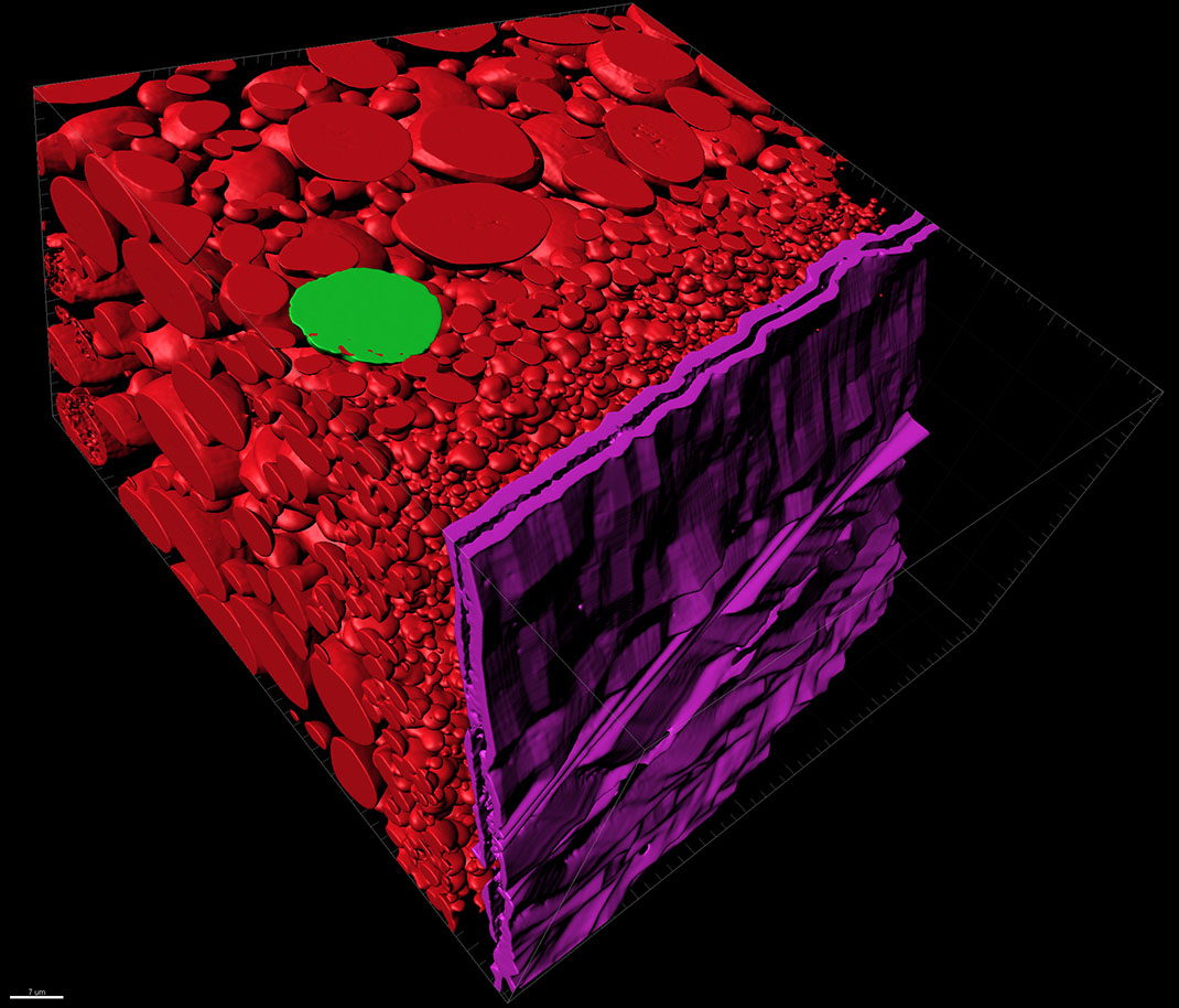
Scattered across this image are segmented cross sections of a clawed frog egg. The image was taken with a Zeiss Sigma VP 3View Serial Block-Face SEM (SBF-SEM), segmented in Microscopy Image Browser and imported into Imaris to create a surface. The goal of the segmentation was to measure the individual volumes of the different sized yolk. The large red circles mark the yolk substance that feeds the developing embryo. The violet segmented structure represents the two-layered walls of the yolk. Clawed frogs are often used as an experimental model for studies like this one that help us understand how organisms develop.
The Jing Yang Lab is interested in germ plasm in Xenopus oocytes which are organized into small "islands" and dispersed to the vegetal cortex of the oocyte. After fertilization, these small islands come together and form very large aggregates. As the embryo divide, a few cells inherit these large germ plasm aggregates and become primordial germ cells. The Lab is interested in the mechanisms controlling these processes. In oocytes and early embryos, these germ plasms are located vegetally, very close to the cortex of the oocyte or embryo. It contains large amounts of mitochondrial, ER, and insoluble large protein complexes. It can be easily identified by TEM.
The aim of using SBF-SEM was to study how the germ plasm dynamics are regulated at the molecular level. The Lab is currently studying some factors which are likely important for germ plasm dynamics and plans to overexpress or knockdown some genes of interest and study how the manipulation would affect either the localization/organization of germ plasm.