


Illinois Researchers Explore Intersection of Imaging Technology and AI
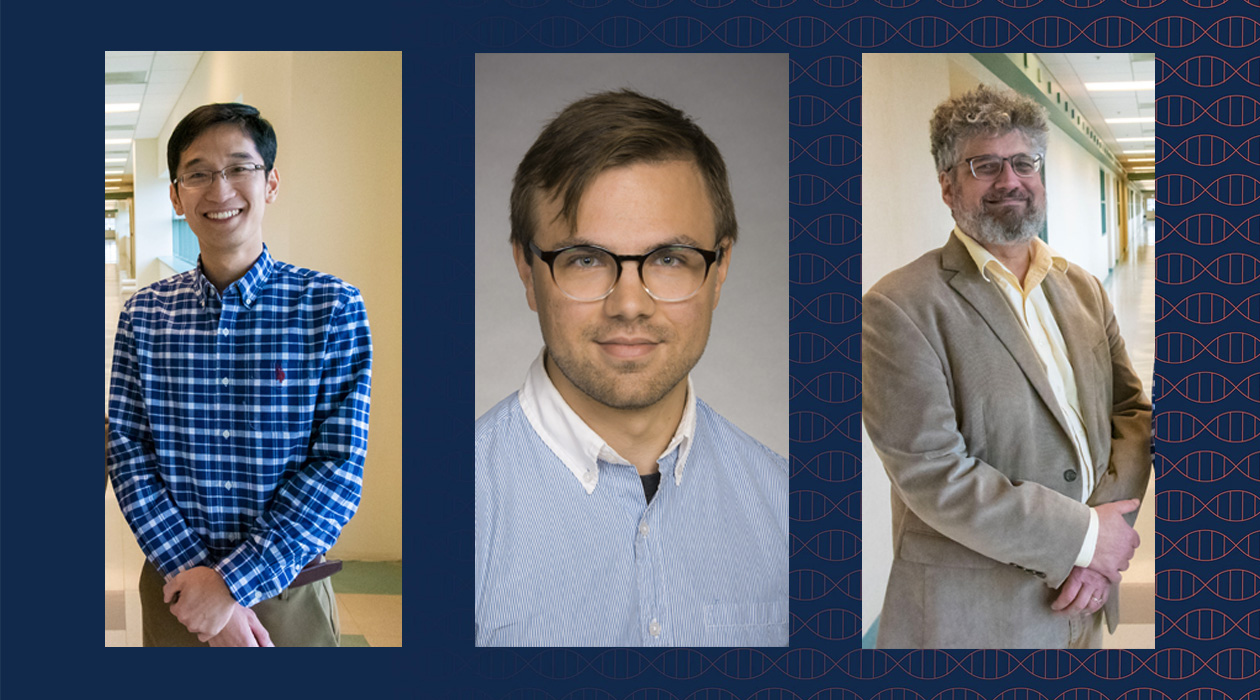
Pictured left to right: bioengineering professor Fan Lam, postdoctoral researcher Timothy Trinklein, and chemistry professor Jonathan Sweedler / Jenna Kurtzweil

Share
Understanding precisely what molecules are inside the body and how they are localized is not only an important scientific pursuit but also valuable for disease diagnosis and treatment. Mass spectrometry imaging (MSI) is an especially potent tool. It works by taking sections of tissue and dividing them into tens of thousands or even millions of individual pixels. This allows researchers to locate metabolites, lipids, peptides, proteins, and other molecules, providing a huge amount of useful data for studying and understanding biochemistry in health and disease.
That quantity of data is MSI’s strength, but it can also be a challenge. There is so much information that is difficult to parse effectively and MSI is relatively slow compared to other imaging techniques. But, according to a review led by postdoctoral researcher Timothy Trinklein, bioengineering professor Fan Lam (GNDP), chemistry professor Jonathan Sweedler (BSD/CAMBERS/MMG) and their team of researchers, artificial intelligence may provide a solution. Their review outlining the potential for the integration of these technologies has been published in Chemical Reviews, an ACS publication.
One of the central opportunities for integrating these technologies lies in accelerating data acquisition. While MSI is extremely thorough, that very precision—rendering thousands to millions of pixels to generate a large number of high-resolution molecular maps—can take hours to even weeks. Lam et al. have already demonstrated, in a paper recently published to Nature Methods, that deep learning can accelerate this process significantly. Lam explains: “One can also ‘adaptively’ accelerate the instrument, using information obtained from the first few pixels to decide where to guide the instrument to maximize information gain. With these enabling AI-driven acceleration technologies, we think it will be possible to apply MSI to large cohorts, clinical samples, or even 3D imaging of entire organs, in the near future.”
Speed is not the only area where AI can enhance MSI. Since the data sets in MSI are so huge, visualizing and interpreting those enormous amounts of information can be uniquely challenging. The group covers how AI can be helpful for reviewing these large data sets by making the data easier for researchers to interpret through techniques like dimensionality reduction, which transforms large data sets into smaller (and more informative) representations to discover unique structures in the data.
AI can even be useful in combining MSI with other imaging technologies, enhancing the capabilities of each. “We are seeing an increasing effort to combine conventional fluorescence or brightfield microscopy with MSI, to provide a morphological reference to the chemical information from MSI,” said Lam and Trinklein. However, this is just the tip of the iceberg. Other techniques that AI could combine with MSI include MRI, vibrational spectroscopy, or even electron microscopy. One combination the team is particularly excited about is virtual staining. Lam explains: “Virtual staining is a process in which AI predicts a conventional histological stain from MSI data. This technique is becoming increasingly common in histology labs to predict stains from label-free optical images and is also being applied to other imaging methods like vibrational imaging from bioengineering professor Rohit Bhargava’s (CGD) group here at Illinois. Virtual MSI, once more fully developed, should save substantial time and effort.”
Though the potential of AI in MSI is exciting, Lam also advises caution. The technology is not yet at a place where researchers can simply input MSI data and produce new biological insights. The data still has to be checked by experts in whichever domain the technology is being applied. “AI can create convincing data that are not supported by the measurements,” cautions the research team. “Successful MSI studies still require a strong experimental approach, and significant time in the lab to sample tissue sections, prepare them for analysis, and run the instrumentation. While MSI has been around for decades, there are still many aspects of the sample preparation and ionization processes that are not well understood and cannot be modeled, and so require trial and error and expert intuition.”
The team hopes that this review can bridge the fields of MSI and AI, and encourage those who are experts in one area or the other to see the power of integration. They also hope that this review can encourage industry partners to adapt AI into their workflows by accelerating data acquisition, compressing data for faster transfer, and incorporating modern analysis tools into commercial packages.
The future for the integration of AI and MSI is bright. Speeding up data acquisition and enabling more efficient data handling will go a long way in making this powerful imaging technology more accessible. “With the advances in physical AI and communication protocols coming out of leading AI labs and companies that allow AI models to interact with physical instruments, there is a tremendous opportunity to make MSI more ‘intelligent’ and studies more automatic,” said Lam. “We hope that, as some of these tools mature, the broader MSI field will reach a consensus on how to best tackle some of these goals with AI.”
The breakthroughs have already begun. The first author of this paper, postdoctoral researcher Timothy Trinklein, has just received an NIH K99/R00 award for a project which integrates AI-powered computational methods and cutting-edge MSI technologies to push the boundaries of neurobiochemical imaging.