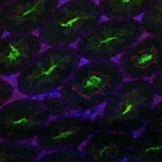
A cross section of mouse testis showing germ cells and spermatogenesis. The green color is basigin expression, which is a cell membrane protein. The red color is claudin-4 expression, which is an epithelial tight junction protein. The blue color is DAPI staining of nuclei. The picture is taken on confocal microscope Zeiss LSM 710. The tubule structures are seminiferous tubules, and that is where spermatogenesis occurs. In each seminiferous tubule, there are 4-6 layers of germs cells at different developmental stages. In the lumen, the bright green color represents the tails of spermatozoa. The red colors in the lumen represent mature spermatozoa. The interstitial cells between each seminiferous tubule are Leydig cells and they produce male sex hormone testosterone, which is essential for normal spermatogenesis and maintaining male characteristics.
