


Bioengineering Lab Develops Innovative Method of Macrophage Imaging
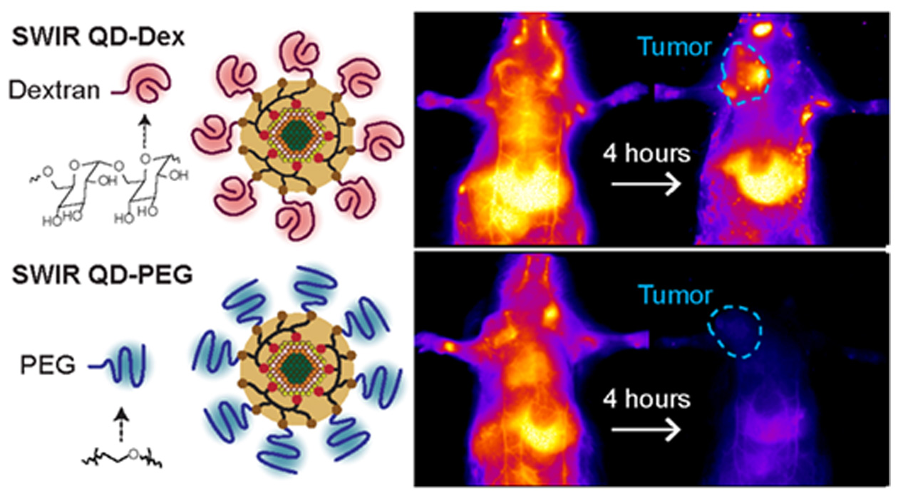
The visual abstract from the paper, entitled “Macrophage-Targeted Quantum Dots with Short-Wave Infrared Emission for In Vivo Breast Cancer Imaging,” published in Bioconjugate Chemistry.

Share
Immune cells are thought to play a major role in whether tumors respond to therapies, but there is no reliable way to monitor these cells in a tumor routinely over time during therapy. Here Illinois investigators use quantum dots that emit short-wave-infrared light, which allows macrophage immune cells to be monitored within tumors.
Understanding macrophages is one of the most important battles in the fight against cancer. As part of the immune system, macrophages can provide researchers with valuable information about a tumor. Under the right conditions, they can help suppress and even kill cancer cells. However, depending on the phenotype of the macrophage, these cells can work against the body and may even protect the tumor. Knowing how macrophages are behaving can provide clinicians with vital information about how responsive a particular cancer may be to a particular treatment. Bioengineering professor Andrew Smith explains: “Too many macrophages and in the wrong phenotype can make tumors ‘immunologically cold’ and nonresponsive.”
A new study from the Department of Bioengineering at The Grainger College of Engineering provides a novel method for tracking these cells. Published in Bioconjugate Chemistry by first author and bioengineering PhD student Fengyuan Xu in the lab of Donald Biggar Willett Faculty Scholar and Professor of Bioengineering Andrew Smith (CGD), the technique utilizes quantum dots that emit infrared light. The dots are coated in dextran, a molecule that macrophages are prone to consume. When introduced to tumors, macrophages pick up the quantum dots as they continue to glow with infrared light. This allows researchers to image the cells and monitor their behavior. “We hope this imaging agent can give us the ability to assess how individual tumors respond to agents which reactivate the immunological state to one that is ‘hot,’” said Smith.
Using quantum dots that emit infrared light is simple and requires less infrastructure. “Only a simple camera system and LED light source are needed with minimal training,” said Smith. “Compared with PET, SPECT, and MRI, which require complex and expensive infrastructure, imaging can be applied rapidly and repeatedly with little cost or time.”
Next, the team hopes to evaluate how well the quantum dots predict the effects of immunotherapy on tumors, including macrophage movement, changes in macrophage phenotypes, and whether quantum dot imaging can predict survival. In the future, Smith hopes this technology can improve testing of cancer therapeutics. “We hope the imaging agents we developed will reduce the burden of animal studies,” explains Smith, “and allow greater depth of understanding of how macrophages in tumors respond to therapies. This may expedite the development and translation of new immunotherapies to the clinic.”
The project was supported by the Cancer Center at Illinois (CCIL), a National Cancer Institute-designated Basic Cancer Center.