Serial Block Face Scanning Electron Microscope (SBF-SEM)
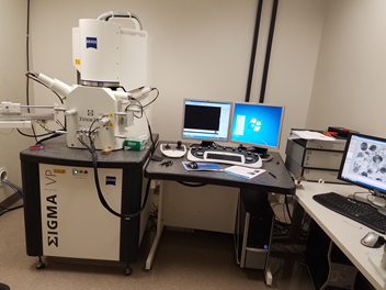
The SBF-SEM is an automated technique for obtaining serial block face images and 3D data using a SEM. Our Zeiss Sigma VP 3View system is a Gatan 3View SBF microtome housed in a Gemini SEM column. The microtome is equipped with a diamond knife which is mounted inside the chamber of the SEM and shaves of <50nm (15 – 200nm) of the sample in between imaging. Images are collected using a backscattered (BD) electron detector which results in a TEM-like image. The SBF-SEM imaging process is fully automated, allowing for a large volume acquisition without the risk of losing sections.
The instrument has the following features and advantages:
This Zeiss Sigma VP 3View system is a Gatan 3View SBF microtome housed in a Gemini SEM column.
It is equipped with several detectors for both regular SEM imaging and SBF imaging. It includes an in-lens detector for detecting secondary (SE) and/or backscattered (BSE) electrons as well as a Variable Pressure Secondary Electron (VPSE G3) detector.
The SMART SEM software is for imaging in SEM mode and the Gatan Microscopy Suite (also known as Gatan Digital Micrograph) is for SBF imaging and data analysis.
<50nm X, Y stage repeatability allows multi-region imaging without losing data due to imprecise stage motion.
You can automatically acquire images across multiple regions of the sample utilizing multi-regions of interest (mROIs).
Large format image support (up to 32k x 24k) allows very large images to be collected.
15nm Z section thickness without the need to unblur multi-kV images
High performance back-scatter detector allows high speed imaging at low kV without giving up image quality
Links:
Zeiss Sigma 3View Introduction Video: https://www.youtube.com/watch?v=3yk3rbgu49o
Gatan 3View Technical Notes and Protocols: http://www.gatan.com/products/sem-imaging-spectroscopy/3view-system
Zeiss Sigma 3View White Paper: http://applications.zeiss.com/C125792900358A3F/0/12129D5013398101C1257A91004312DF/
Zeiss Sigma 3View Webinar: https://www.zeiss.com/microscopy/us/products/scanning-electron-microscopes/merlin-life-science.html
3View Sample Processing Request Form:
Manufacturer: Zeiss
Equipment Model: Sigma VP
Location: Room 15, Carl R. Woese Institute for Genomic Biology
Contact Person: Kingsley Boateng, 217-333-1642, kboateng@igb.illinois.edu
Sample Images and Videos:
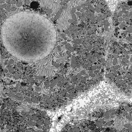
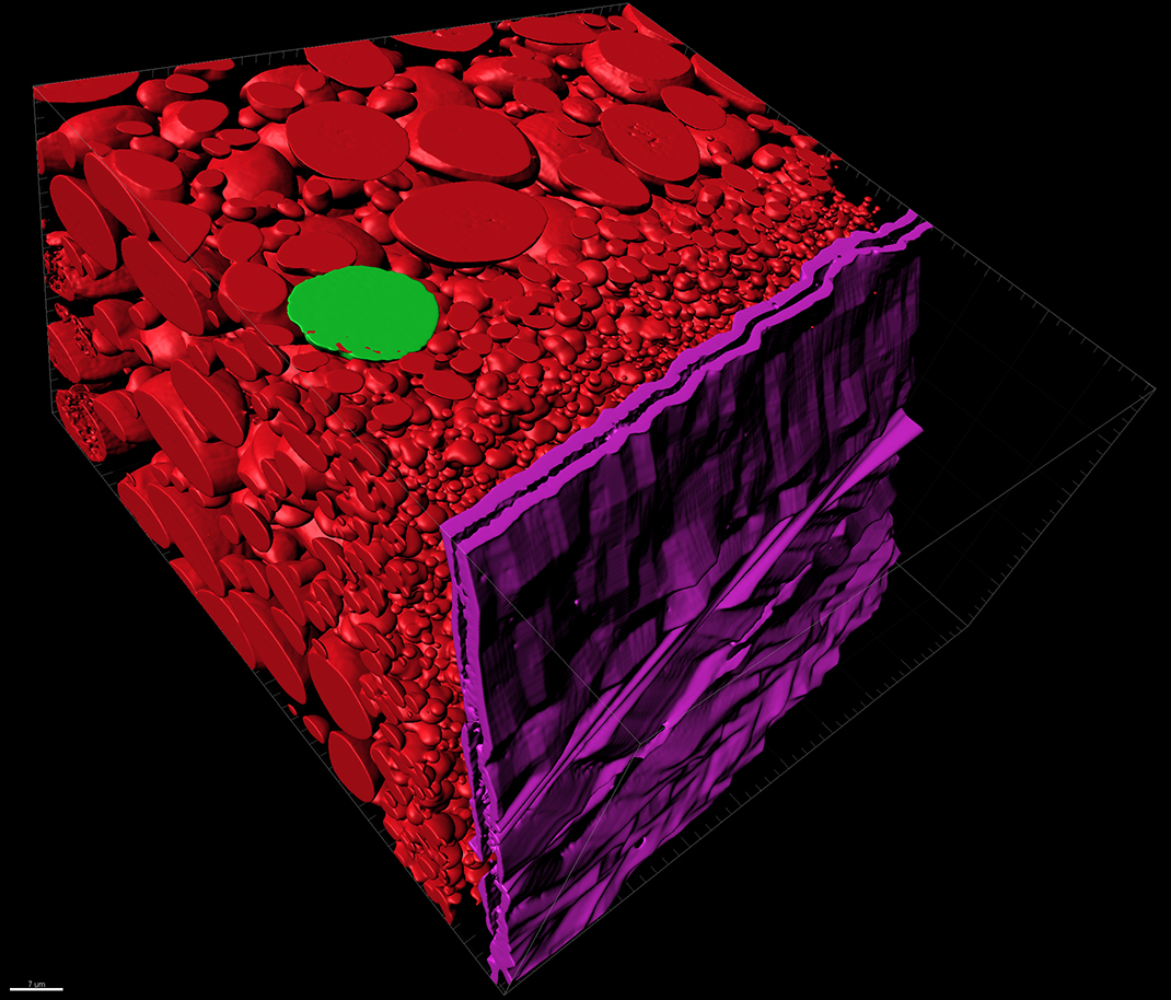
Liver Tissue
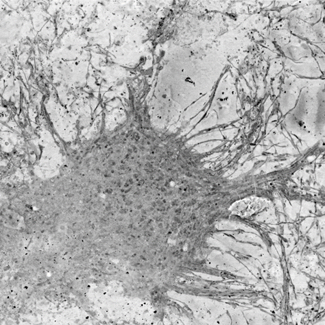
Neurons
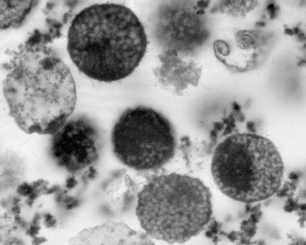
Archaea cells
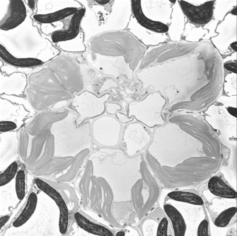
Plant leaf - chloroplast
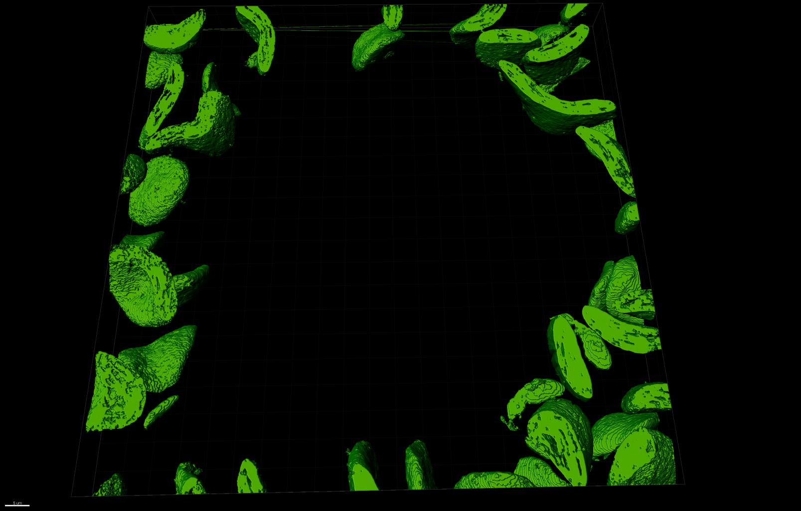
Segmented chloroplast with MIB Software
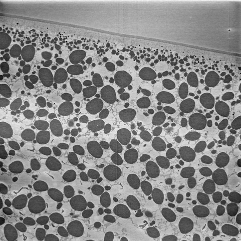
Xenopus laevis oocytes (frog egg yoke)
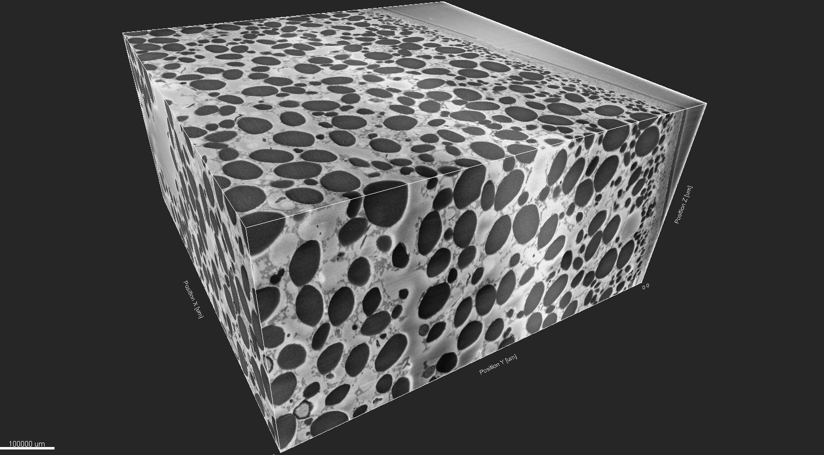
Segmented frog egg-yoke with MIB Software