New photonic crystal approach can enable sensitive and affordable detection of biomarkers

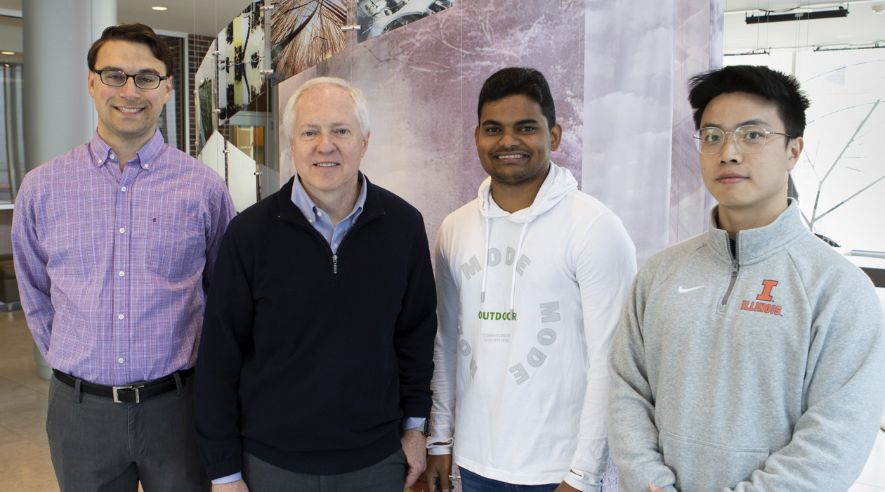
The authors of the study, from left to right: Joseph Tibbs, Brian Cunningham, Seemesh Bhaskar and Weinan Liu.
Biomarkers are small molecules of interest to researchers, because they can indicate underlying diseases, often even before symptoms even appear. However, detecting these markers can be challenging as they are often present in very low quantities, especially in the early stages of a disease. Traditional detection methods, while effective, usually require expensive components like prisms, metal films, or optical objectives.
In a recent paper published in Applied Physics Letters, researchers at the University of Illinois Urbana-Champaign have unveiled a novel approach to detecting low concentrations of biomarkers that paves the way for biodetection technology that is simple to use, highly sensitive, and surprisingly affordable.
“The goal of this technology is early diagnostics, to be able to detect molecules associated with diseases at very low concentrations, sometimes a few molecules per millions, very early on,” said Seemesh Bhaskar, a postdoctoral researcher in the Cunningham lab and first author on the study. “Looking for very small concentrations of micro-RNA, circulating tumor DNA, and exosomes, for example, can help determine whether a patient will develop cancer one or two years down the line.”
Early detection of biomarkers is crucial for predicting and managing diseases effectively. There are many strategies for measuring the presence and concentration of biomarkers, but a common approach involves binding them with a fluorescent molecule, called a fluorophore, which emits fluorescence when excited with light.
Bhaskar noted that while there are technologies adept at detecting these low levels of fluorescent biomarkers, they are often bulky and expensive, limiting their accessibility in healthcare, particularly in resource-limited areas.
The approach encompasses a novel phenomenon for detecting light, called radiating guided mode resonance, which utilizes photonic crystals — thin pieces of glass with small gratings on the surface. These gratings help direct the photons, which are small particles of light, emitted from biomarkers along a pathway via a steering effect. This pathway is “tuned” to match the wavelength of the fluorescence emitted by the biomarkers, optimizing light collection and enhancing detection sensitivity.
Bhaskar likens this to a rhythmic dance of light energy within the crystal, where light is amplified while taking on the properties of the photonic crystal. One property of the crystal, called polarization selection, equalizes the polarization of the light, making for clearer and sharper detection of fluorescence. Together, this can result in an output that is 100 times stronger.
“To me, this is a whole new way of looking into the properties of light itself,” said Bhaskar. “The photons adapt, change, and evolve as they pass through the photonic crystal. The light picks up new characteristics without losing its essence. It's a testament to the adaptability and transformative power of light.”
Discovery of this new phenomenon sets the stage for future detection platforms that will be able to detect molecules at picomolar levels without relying on costly components, making biodetection technology more sensitive, accessible, and affordable.
While radiating guided mode resonance can theoretically be used to enhance detection of many different biomarkers, the Cunningham lab is particularly interested in early cancer detection. The new phenomenon holds promise for affordable technology that will be critical for populations in resource-limited settings, where early disease detection and treatment can make a profound difference.
One of the lab’s long-term goals after development of this technology is to make it compatible with smartphones, further enhancing its accessibility. The envisioned future product would be a simple fixture attached to a smartphone’s camera, allowing a photonic crystal to illuminate a test sample while the phone’s camera measures the fluorescence emission.
“We are creating biosensing systems that are extremely sensitive while utilizing simple and inexpensive detection instruments,” said Brian Cunningham (CGD leader/MMG), a professor of electrical and computer engineering and program leader at the Cancer Center at Illinois. “This is what creates a path toward sophisticated health diagnostics making their way to our health clinics, farms, and homes.”
The study was funded by NIH, NSF, and the Cancer Center at Illinois. The paper can be found at https://doi.org/10.1063/5.0203999
