Damaged liver cells undergo reprogramming to regenerate
Share
In Greek mythology, Zeus punishes the trickster Prometheus by chaining him to a rock and sending an eagle to eat a portion of his liver every day, in perpetuity. It was the right organ to target – the liver has the ability to regenerate itself, though not overnight nor for eternity.
New research conducted by biochemists at the University of Illinois has determined how damaged liver cells repair and restore themselves through a signal to return to an early stage of postnatal organ development. The findings are reported in the journal Nature Structural & Molecular Biology.
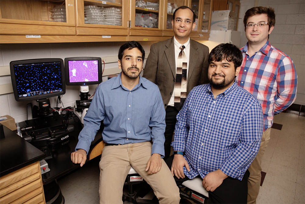
“The liver is a resilient organ,” said biochemistry professor and IGB member in the Omics Nanotechnology for Cancer Precision Medicine and Gene Networks in Neural & Developmental Plasticity research themes Auinash Kalsotra, who led the new research. “It can restore up to 70 percent of lost mass and function after just a few weeks.
“We know that in a healthy adult liver, the cells are dormant and rarely undergo cell division,” he said. “However, if the liver is damaged, the liver cells re-enter the cell cycle to divide and produce more of themselves.”
The human liver can become chronically damaged by toxins such as alcohol and even certain medicines, but still continue to function and self-repair, Kalsotra said.
“This research looked at what is happening at the molecular level in a damaged liver that enables it to regenerate while still performing normal functions,” he said.
Using a mouse model of a liver severely damaged by toxins, the researchers compared injured adult liver cells with healthy cells present during a stage of development just after birth. They found that injured cells undergo a partial reprograming that returns them to a neonatal state of gene expression.
The team discovered that fragments of messenger RNA, the molecular blueprints for proteins, are rearranged and processed in regenerating liver cells in a manner reminiscent of the neonatal period of development. This phenomenon is regulated through alternative splicing, a process wherein exons (expressed regions of genes) are cut from introns (intervening regions) and stitched together in various combinations to direct the synthesis of many different proteins from a single gene. These proteins can have different cellular functions or properties.
“We found that the liver cells after birth use a specific RNA-binding protein called ESRP2 to generate the right assortment of alternatively spliced RNAs that can produce the protein products necessary for meeting the functional demands of the adult liver,” said graduate student Sushant Bangru, the lead author of the study. “When damaged, the liver cells lower the quantity of ESRP2 protein. This reactivates fetal RNA splicing in what is called the ‘Hippo signaling pathway,’ giving it instructions about how to restore and repopulate the liver with new and healthy cells.”
Kalsotra described the science in mythological terms: “When Zeus’ eagle comes in for its daily snack, damaging the liver, the alternatively spliced form of Hippos come into play – repairing Prometheus’s liver so the poor guy can go through this whole punishment again the next day.”
The National Institutes of Health, March of Dimes and American Heart Association supported this research.