Outreach & Public Engagement


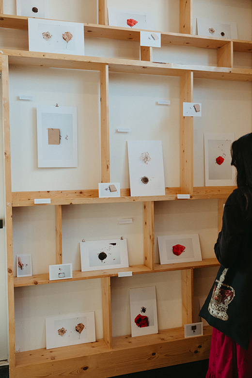
The IGB’s Art of Science program is a celebration of common ground between science and art. Each exhibit comprises images from IGB’s research portfolio, enhanced to highlight the beauty and fascination encountered daily in scientific endeavors.
Read More About Art of Science
Art of Science 16
Perfusion
Julia Pollack
-
Analog Wine Library 129 N Race St. Urbana, IL
Scientist Collaborator
Joseph Tibbs
Instrument
Matlab
Funding Agency
Funded by the National Institutes of Health, Illinois Distinguished Fellowship
Original Imaging
Special Thanks
Nelson family and BodyWork Associates; Matt Cho and Analog Wine Library
Image Rights
Images not for public use without permission from the Carl R. Woese Institute for Genomic Biology
Scientist Collaborator
Bishal Tamang, Gregory Bernard, Sebastian Valera, and Andrew Leakey
Lisa Ainsworth Group
Instrument
Satellite and Unmanned Aerial Vehicle Images
Funding Agency
Bill & Melinda Gates Foundation, Gates Ag One
Original Imaging
Special Thanks
Nelson family and BodyWork Associates; Matt Cho and Analog Wine Library
Image Rights
Images not for public use without permission from the Carl R. Woese Institute for Genomic Biology
Scientist Collaborator
Jeanaflor Crystal T. Concepcion
Lisa Ainsworth Group
Instrument
Canon 5D
Funding Agency
Bill & Melinda Gates Foundation, Gates Ag One
Original Imaging
Special Thanks
Nelson family and BodyWork Associates; Matt Cho and Analog Wine Library
Image Rights
Images not for public use without permission from the Carl R. Woese Institute for Genomic Biology
Scientist Collaborator
Jeanaflor Crystal T. Concepcion
Lisa Ainsworth Group
Instrument
Canon 5D
Funding Agency
Bill & Melinda Gates Foundation, Gates Ag One
Original Imaging
Special Thanks
Nelson family and BodyWork Associates; Matt Cho and Analog Wine Library
Image Rights
Images not for public use without permission from the Carl R. Woese Institute for Genomic Biology
Scientist Collaborator
Jeanaflor Crystal T. Concepcion
Lisa Ainsworth Group
Instrument
Canon 5D
Funding Agency
Bill & Melinda Gates Foundation, Gates Ag One
Original Imaging
Special Thanks
Nelson family and BodyWork Associates; Matt Cho and Analog Wine Library
Image Rights
Images not for public use without permission from the Carl R. Woese Institute for Genomic Biology
Scientist Collaborator
Danny Ryerson, IGB Outreach Group
Instrument
Zeiss Axioscope
Funding Agency
Carl R. Woese Institute for Genomic Biology
Original Imaging
Special Thanks
Nelson family and BodyWork Associates; Matt Cho and Analog Wine Library
Image Rights
Images not for public use without permission from the Carl R. Woese Institute for Genomic Biology
Scientist Collaborator
Danny Ryerson, IGB Outreach Group
Instrument
Zeiss Axioscope
Funding Agency
Carl R. Woese Institute for Genomic Biology
Original Imaging
Special Thanks
Nelson family and BodyWork Associates; Matt Cho and Analog Wine Library
Image Rights
Images not for public use without permission from the Carl R. Woese Institute for Genomic Biology
Scientist Collaborator
Danny Ryerson, IGB Outreach Group
Instrument
Zeiss Axioscope
Funding Agency
Carl R. Woese Institute for Genomic Biology
Original Imaging
Special Thanks
Nelson family and BodyWork Associates; Matt Cho and Analog Wine Library
Image Rights
Images not for public use without permission from the Carl R. Woese Institute for Genomic Biology
Scientist Collaborator
Danny Ryerson, IGB Outreach Group
Instrument
Zeiss Axioscope
Funding Agency
Carl R. Woese Institute for Genomic Biology
Original Imaging
Special Thanks
Nelson family and BodyWork Associates; Matt Cho and Analog Wine Library
Image Rights
Images not for public use without permission from the Carl R. Woese Institute for Genomic Biology
Scientist Collaborator
Hannah Theriault, Brendan Harley Group
Instrument
Multiphoton Confocal Microscope Zeiss 710 with Mai Tai eHP Ti:sapphire laser, 5x and 20x
Funding Agency
National Institutes of Health
Original Imaging
Special Thanks
Nelson family and BodyWork Associates; Matt Cho and Analog Wine Library
Image Rights
Images not for public use without permission from the Carl R. Woese Institute for Genomic Biology
Scientist Collaborator
Hannah Theriault, Brendan Harley Group
Instrument
Multiphoton Confocal Microscope Zeiss 710 with Mai Tai eHP Ti:sapphire laser, 5x and 20x
Funding Agency
National Institutes of Health
Original Imaging
Special Thanks
Nelson family and BodyWork Associates; Matt Cho and Analog Wine Library
Image Rights
Images not for public use without permission from the Carl R. Woese Institute for Genomic Biology
Scientist Collaborator
Hannah Theriault, Brendan Harley Group
Instrument
Multiphoton Confocal Microscope Zeiss 710 with Mai Tai eHP Ti:sapphire laser, 5x and 20x
Funding Agency
National Institutes of Health
Original Imaging
Special Thanks
Nelson family and BodyWork Associates; Matt Cho and Analog Wine Library
Image Rights
Images not for public use without permission from the Carl R. Woese Institute for Genomic Biology
Scientist Collaborator
Jayita De, Pratik Banerjee Group
Instrument
Zeiss LSM 880 Airyscan
Funding Agency
United States Department of Agriculture, University of Illinois Urbana-Champaign
Original Imaging
Special Thanks
Nelson family and BodyWork Associates; Matt Cho and Analog Wine Library
Image Rights
Images not for public use without permission from the Carl R. Woese Institute for Genomic Biology
Scientist Collaborator
Kevin Neumann, Alison Bell Group; Andy Suarez Group
Instrument
Canon 5D
Funding Agency
University of Illinois Urbana-Champaign
Special Thanks
Nelson family and BodyWork Associates; Matt Cho and Analog Wine Library
Image Rights
Images not for public use without permission from the Carl R. Woese Institute for Genomic Biology
Scientist Collaborator
Gunnar Thompson, Brendan Harley Group
Instrument
Zeiss LSM 710 Confocal Microscope
Funding Agency
National Institutes of Health
Original Imaging
Special Thanks
Nelson family and BodyWork Associates; Matt Cho and Analog Wine Library
Image Rights
Images not for public use without permission from the Carl R. Woese Institute for Genomic Biology
Scientist Collaborator
Duncan Nall and Austin Cyphersmith, Andrew Smith Group
Instrument
Zeiss Lightsheet Z1 Microscope
Funding Agency
Cancer Center at Illinois
Original Imaging
Special Thanks
Nelson family and BodyWork Associates; Matt Cho and Analog Wine Library
Image Rights
Images not for public use without permission from the Carl R. Woese Institute for Genomic Biology
Scientist Collaborator
Quang Nguyên, Martha Gillette Group
Instrument
LSM 900 Confocal Microscope
Funding Agency
National Institutes of Health
Original Imaging
Special Thanks
Nelson family and BodyWork Associates; Matt Cho and Analog Wine Library
Image Rights
Images not for public use without permission from the Carl R. Woese Institute for Genomic Biology
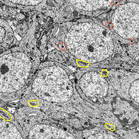
Scientist Collaborator
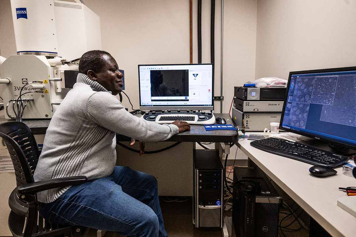
Vipendra Kumar, Nien-Pei Tsai Group
Instrument
Zeiss Sigma VP 3View Serial Block-Face Scanning Electron Microscope
Original Imaging
Special Thanks
Nelson family and BodyWork Associates; Matt Cho and Analog Wine Library
Image Rights
Images not for public use without permission from the Carl R. Woese Institute for Genomic Biology
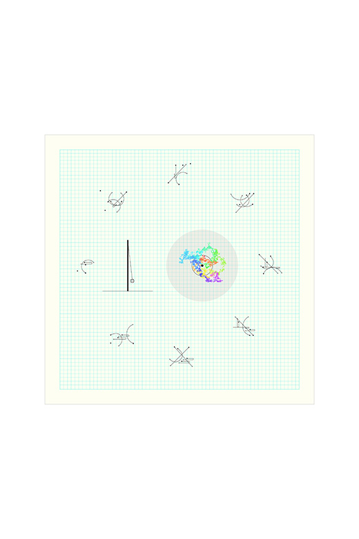
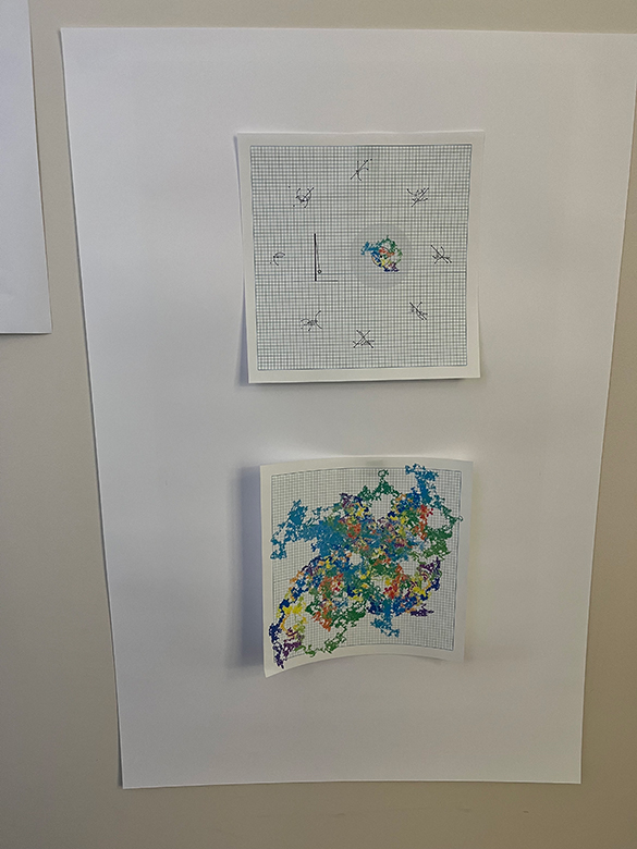
ALEATORIC PLAYGROUND
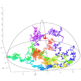
Life is built from a complex web of microscopic processes. In less than a second, innumerable biological events are occurring, far beneath the limits of the human eye. Modern technology provides a lens to view these miniscule operations, but small things can be difficult to distinguish from each other, and fast processes still evade our observation. Better understanding of the nanoscale and new detection tools can be harnessed to improve human health.
The image depicts the trajectory of a gold nanoparticle, bouncing around in a microscopic game of tetherball. Anchored to a central point by a tiny piece of DNA, the particle is constrained in space, simplifying this computer-simulated system to a game of randomness and probabilities. Following the rainbow path, jumpy and jagged as it collides with billions of water molecules, we can track the nanoparticle’s movement in time—from red to purple in mere microseconds. By analyzing the statistical properties of these fluctuations, the researchers can learn things about the particle and the space it occupies. Understanding these micro- scale dynamics is a step towards better detection of small things: a strand of DNA, a single virus, a molecular marker of early cancer growth.
BETWEEN THESE TWO POLES
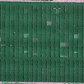
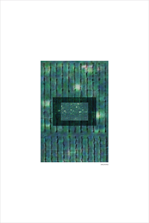
Across the midwest, a variegated and slightly irregular grid of fields stretches like a green rag rug. Flying into Urbana-Champaign, Illinois, it is easy to see how flush the vegetation is; flush to the flat ground, and flush with life. The corn and soybeans grown here sustain communities beyond our state, reaching regions with scenery that may be more beautiful to some but are less hospitable to staple crops.
Along with the crops themselves, our farmland yields knowledge of how to make them more resilient. Researchers conduct field studies of soybean growth that rely on a combination of labor-intensive data collection and, now, collection and analysis of photographs like these taken by satellites and unmanned aerial vehicles. As the resolution of this type of imagery continues to improve, we can discern individual differences in plant growth from above. Using this technology, researchers can follow and improve the growth of traditional crops in new ways, working toward food security for all.
CHARACTER OF PULSE (1 of 3)
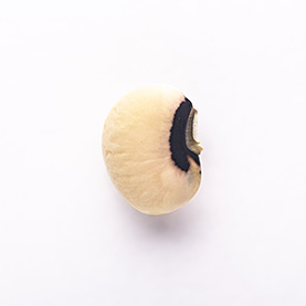
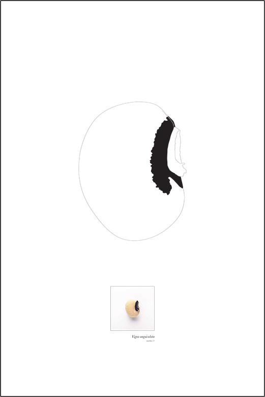
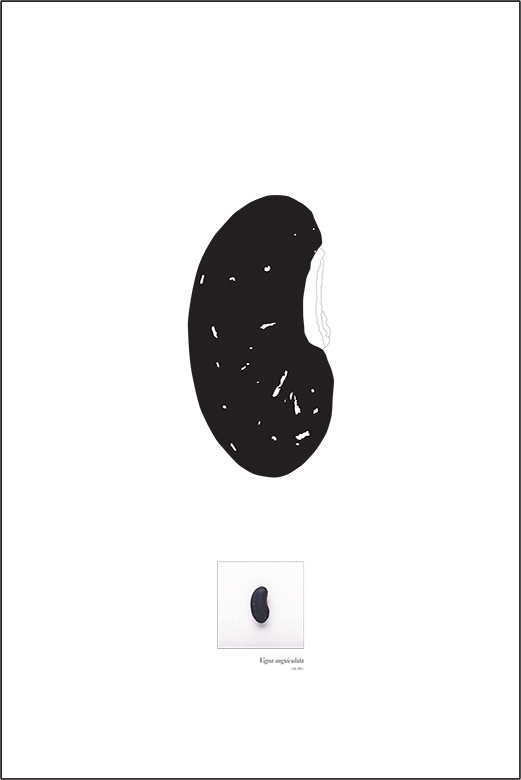
Cowpea, black-eyed pea, crowder-pea, ñebbe—these small and humble-looking beans carry the potential to feed the world. Cowpeas were one of the earliest crops to be domesticated, and were spread from Africa through multiple waves of migration, populating the world with myriad varieties. They are a “hungry-season crop,” nutritious, fast-growing, and tolerant of drought. The mature seed of each variety has its own pigmentation pattern: solid white, black, brown or red, dappled with spots, or featuring the single dark spot that gave rise to one of their names.
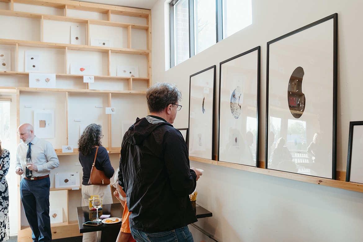
Each cowpea variety also offers a unique set of traits. The varying pigmentation corresponds to slightly different suites of dietary components in each type of bean. The plants themselves also differ, with a range of leaf and canopy sizes and shapes. Researchers are working to document these traits and identify the genes that help shape them to produce more resilient and nutritious varieties. In this way, one of the world’s oldest crops may also become the crop of the future.
CHARACTER OF PULSE (2 of 3)
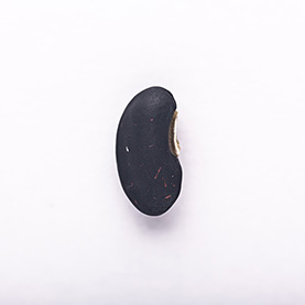
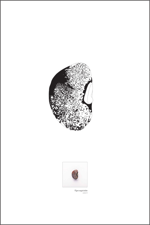
Cowpea, black-eyed pea, crowder-pea, ñebbe—these small and humble-looking beans carry the potential to feed the world. Cowpeas were one of the earliest crops to be domesticated, and were spread from Africa through multiple waves of migration, populating the world with myriad varieties. They are a “hungry-season crop,” nutritious, fast-growing, and tolerant of drought. The mature seed of each variety has its own pigmentation pattern: solid white, black, brown or red, dappled with spots, or featuring the single dark spot that gave rise to one of their names.
Each cowpea variety also offers a unique set of traits. The varying pigmentation corresponds to slightly different suites of dietary components in each type of bean. The plants themselves also differ, with a range of leaf and canopy sizes and shapes. Researchers are working to document these traits and identify the genes that help shape them to produce more resilient and nutritious varieties. In this way, one of the world’s oldest crops may also become the crop of the future.
CHARACTER OF PULSE (3 of 3)
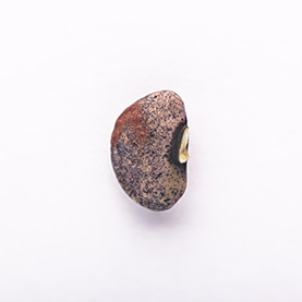
Cowpea, black-eyed pea, crowder-pea, ñebbe—these small and humble-looking beans carry the potential to feed the world. Cowpeas were one of the earliest crops to be domesticated, and were spread from Africa through multiple waves of migration, populating the world with myriad varieties. They are a “hungry-season crop,” nutritious, fast-growing, and tolerant of drought. The mature seed of each variety has its own pigmentation pattern: solid white, black, brown or red, dappled with spots, or featuring the single dark spot that gave rise to one of their names.
Each cowpea variety also offers a unique set of traits. The varying pigmentation corresponds to slightly different suites of dietary components in each type of bean. The plants themselves also differ, with a range of leaf and canopy sizes and shapes. Researchers are working to document these traits and identify the genes that help shape them to produce more resilient and nutritious varieties. In this way, one of the world’s oldest crops may also become the crop of the future.
FABRIC OF NATURE (1 of 4)
Our distant evolutionary ancestors did not contain their blood in a system of branching vessels. Animals with small and simple body plans are able to rely on the physics of diffusion to carry nutrients through their tissues and to rid them of waste. In our invertebrate relatives, hemolymph is stirred by a pumping heart to rush through open spaces inside the body, bathing organs in a tide of sugars, salts, and proteins. In the larger bodies of most vertebrate animals, a more coordinated and efficient type of circulation is needed.
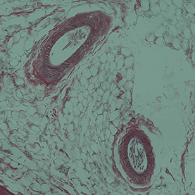
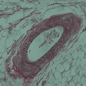
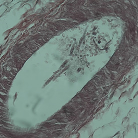
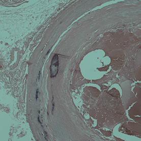
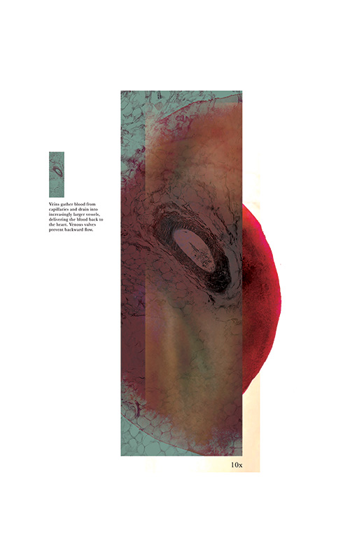
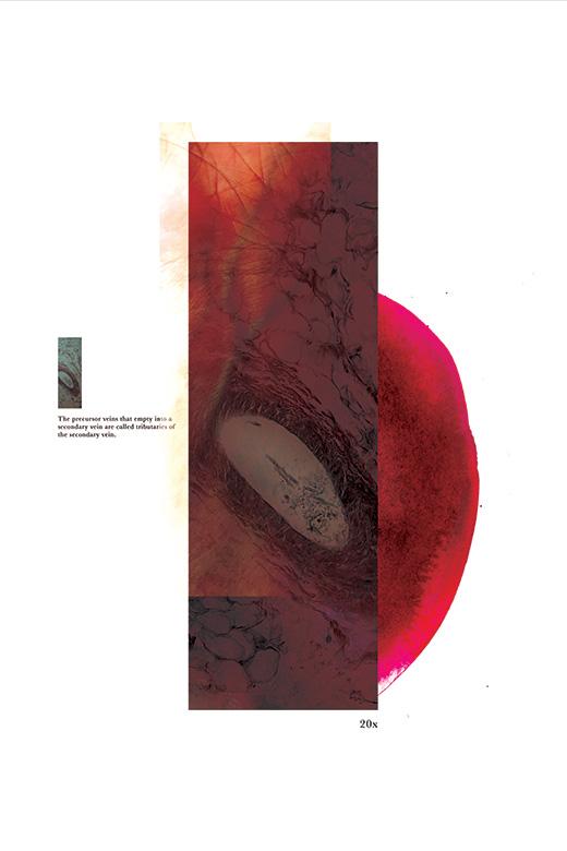
Focusing in on the fine structures of blood vessels allows us to appreciate that they are not simply tubes containing a passive flow of blood. This twinned series of images—a cross-section of an artery and of a vein—directs our attention to the muscle fibers that stretch and contract to push blood toward where it is needed. The artist has contrasted these defined structures with spreading stains of paint, reminding us how quickly blood might escape a vessel’s walls to flow freely once again.
FABRIC OF NATURE (2 of 4)
Our distant evolutionary ancestors did not contain their blood in a system of branching vessels. Animals with small and simple body plans are able to rely on the physics of diffusion to carry nutrients through their tissues and to rid them of waste. In our invertebrate relatives, hemolymph is stirred by a pumping heart to rush through open spaces inside the body, bathing organs in a tide of sugars, salts, and proteins. In the larger bodies of most vertebrate animals, a more coordinated and efficient type of circulation is needed.
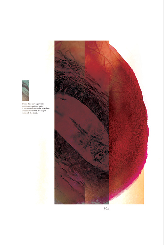
Focusing in on the fine structures of blood vessels allows us to appreciate that they are not simply tubes containing a passive flow of blood. This twinned series of images—a cross-section of an artery and of a vein—directs our attention to the muscle fibers that stretch and contract to push blood toward where it is needed. The artist has contrasted these defined structures with spreading stains of paint, reminding us how quickly blood might escape a vessel’s walls to flow freely once again.
FABRIC OF NATURE (3 of 4)
Our distant evolutionary ancestors did not contain their blood in a system of branching vessels. Animals with small and simple body plans are able to rely on the physics of diffusion to carry nutrients through their tissues and to rid them of waste. In our invertebrate relatives, hemolymph is stirred by a pumping heart to rush through open spaces inside the body, bathing organs in a tide of sugars, salts, and proteins. In the larger bodies of most vertebrate animals, a more coordinated and efficient type of circulation is needed.
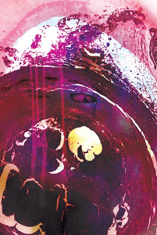
Focusing in on the fine structures of blood vessels allows us to appreciate that they are not simply tubes containing a passive flow of blood. This twinned series of images—a cross-section of an artery and of a vein—directs our attention to the muscle fibers that stretch and contract to push blood toward where it is needed. The artist has contrasted these defined structures with spreading stains of paint, reminding us how quickly blood might escape a vessel’s walls to flow freely once again.
FABRIC OF NATURE (4 of 4)
Our distant evolutionary ancestors did not contain their blood in a system of branching vessels. Animals with small and simple body plans are able to rely on the physics of diffusion to carry nutrients through their tissues and to rid them of waste. In our invertebrate relatives, hemolymph is stirred by a pumping heart to rush through open spaces inside the body, bathing organs in a tide of sugars, salts, and proteins. In the larger bodies of most vertebrate animals, a more coordinated and efficient type of circulation is needed.
Focusing in on the fine structures of blood vessels allows us to appreciate that they are not simply tubes containing a passive flow of blood. This twinned series of images—a cross-section of an artery and of a vein—directs our attention to the muscle fibers that stretch and contract to push blood toward where it is needed. The artist has contrasted these defined structures with spreading stains of paint, reminding us how quickly blood might escape a vessel’s walls to flow freely once again.
GIVE US BACK OUR SUFFERING (1 of 3)
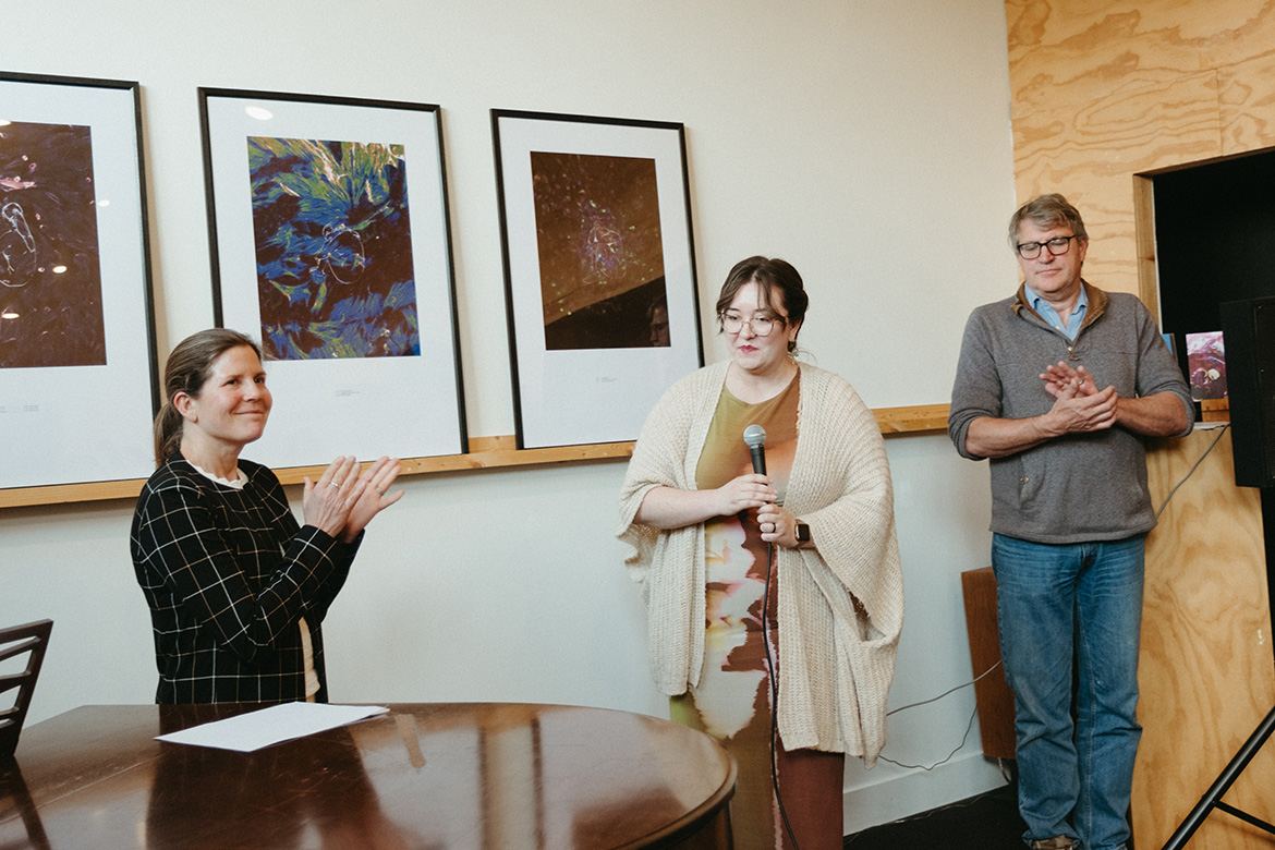
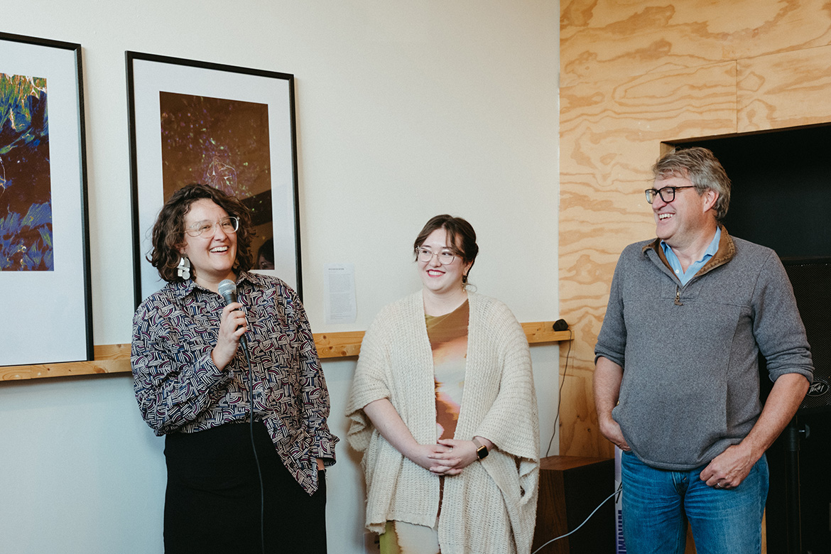
Endometriosis is a common and quietly debilitating disorder, causing chronic pain, fatigue, and a constellation of other possible symptoms. When the inner lining of the uterus, the endometrium, is shed during the menstrual cycle, secreted blood and tissue can occasionally flow upward through the Fallopian tubes and into the abdominal cavity. Scientists have theorized that the disorder begins when endometrial cells carried by menstrual blood manage to implant themselves in other tissues of the abdomen. Their extrauterine growth is the hallmark of the disease, causing inflammation, scarring, and sometimes infertility.
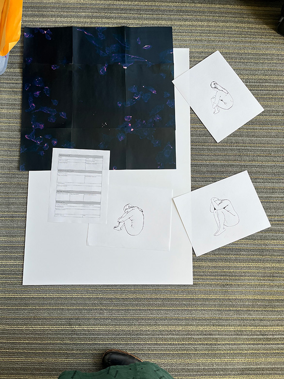
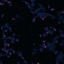
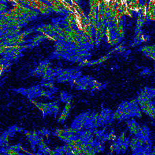
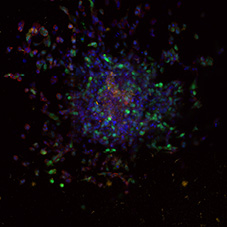
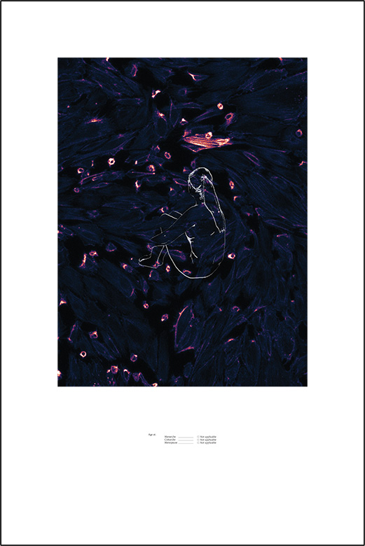
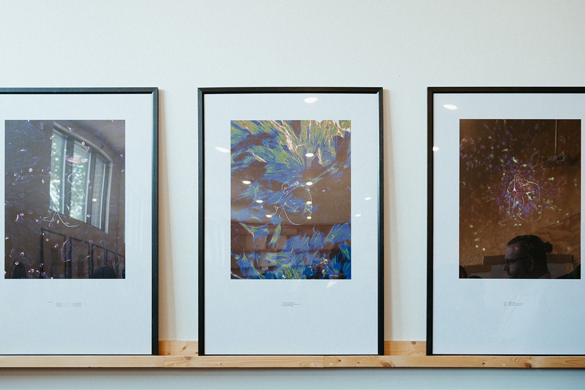
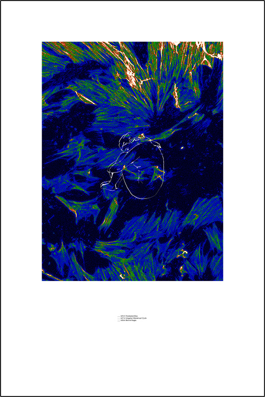
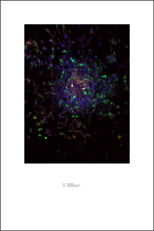
Individuals with endometriosis often wait years to be diagnosed; it is estimated that over half of those affected are never formally diagnosed at all. Their isolated state of limbo has been captured through the line drawings at the center of these works, shadowed by excerpts of the medical forms that cannot contain the totality of their experience. Behind them are images emerging from research that seeks to clarify how endometriosis progresses: orange-labeled ovarian cells, endometriotic disease-causing cells in blue and green, and green-labeled endometriotic cells grown in a spherical gel to track how they might invade a biological structure.
GIVE US BACK OUR SUFFERING (2 of 3)
Endometriosis is a common and quietly debilitating disorder, causing chronic pain, fatigue, and a constellation of other possible symptoms. When the inner lining of the uterus, the endometrium, is shed during the menstrual cycle, secreted blood and tissue can occasionally flow upward through the Fallopian tubes and into the abdominal cavity. Scientists have theorized that the disorder begins when endometrial cells carried by menstrual blood manage to implant themselves in other tissues of the abdomen. Their extrauterine growth is the hallmark of the disease, causing inflammation, scarring, and sometimes infertility.
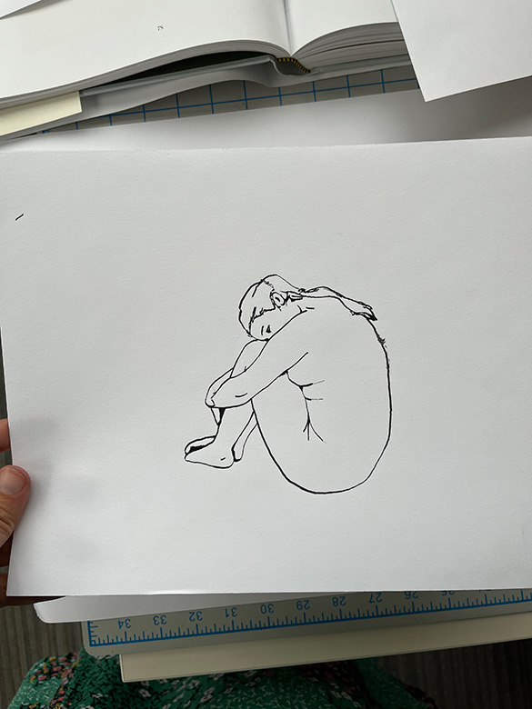
Individuals with endometriosis often wait years to be diagnosed; it is estimated that over half of those affected are never formally diagnosed at all. Their isolated state of limbo has been captured through the line drawings at the center of these works, shadowed by excerpts of the medical forms that cannot contain the totality of their experience. Behind them are images emerging from research that seeks to clarify how endometriosis progresses: orange-labeled ovarian cells, endometriotic disease-causing cells in blue and green, and green-labeled endometriotic cells grown in a spherical gel to track how they might invade a biological structure.
GIVE US BACK OUR SUFFERING (3 of 3)
Endometriosis is a common and quietly debilitating disorder, causing chronic pain, fatigue, and a constellation of other possible symptoms. When the inner lining of the uterus, the endometrium, is shed during the menstrual cycle, secreted blood and tissue can occasionally flow upward through the Fallopian tubes and into the abdominal cavity. Scientists have theorized that the disorder begins when endometrial cells carried by menstrual blood manage to implant themselves in other tissues of the abdomen. Their extrauterine growth is the hallmark of the disease, causing inflammation, scarring, and sometimes infertility.
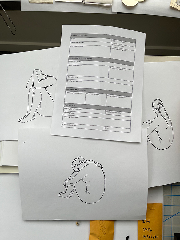
Individuals with endometriosis often wait years to be diagnosed; it is estimated that over half of those affected are never formally diagnosed at all. Their isolated state of limbo has been captured through the line drawings at the center of these works, shadowed by excerpts of the medical forms that cannot contain the totality of their experience. Behind them are images emerging from research that seeks to clarify how endometriosis progresses: orange-labeled ovarian cells, endometriotic disease-causing cells in blue and green, and green-labeled endometriotic cells grown in a spherical gel to track how they might invade a biological structure.
OBJECTS OF MEDICAL TREATMENT
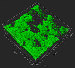
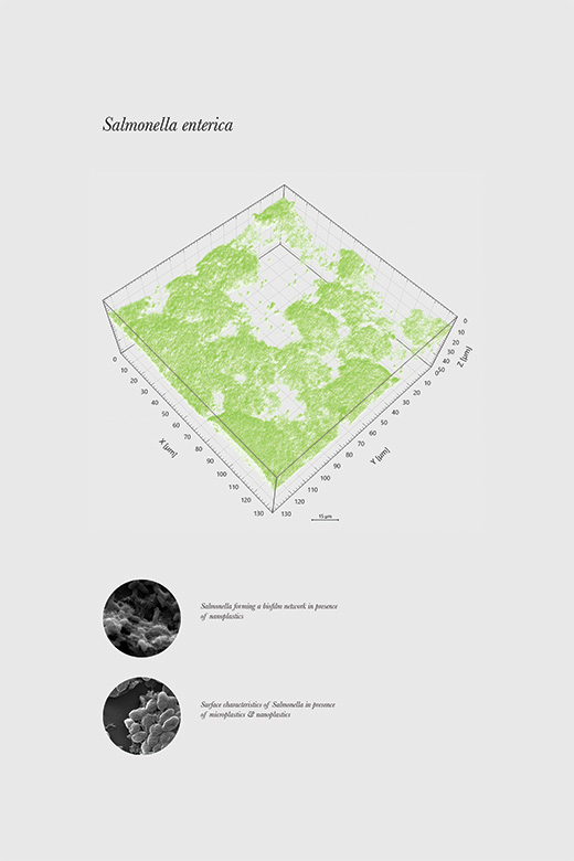
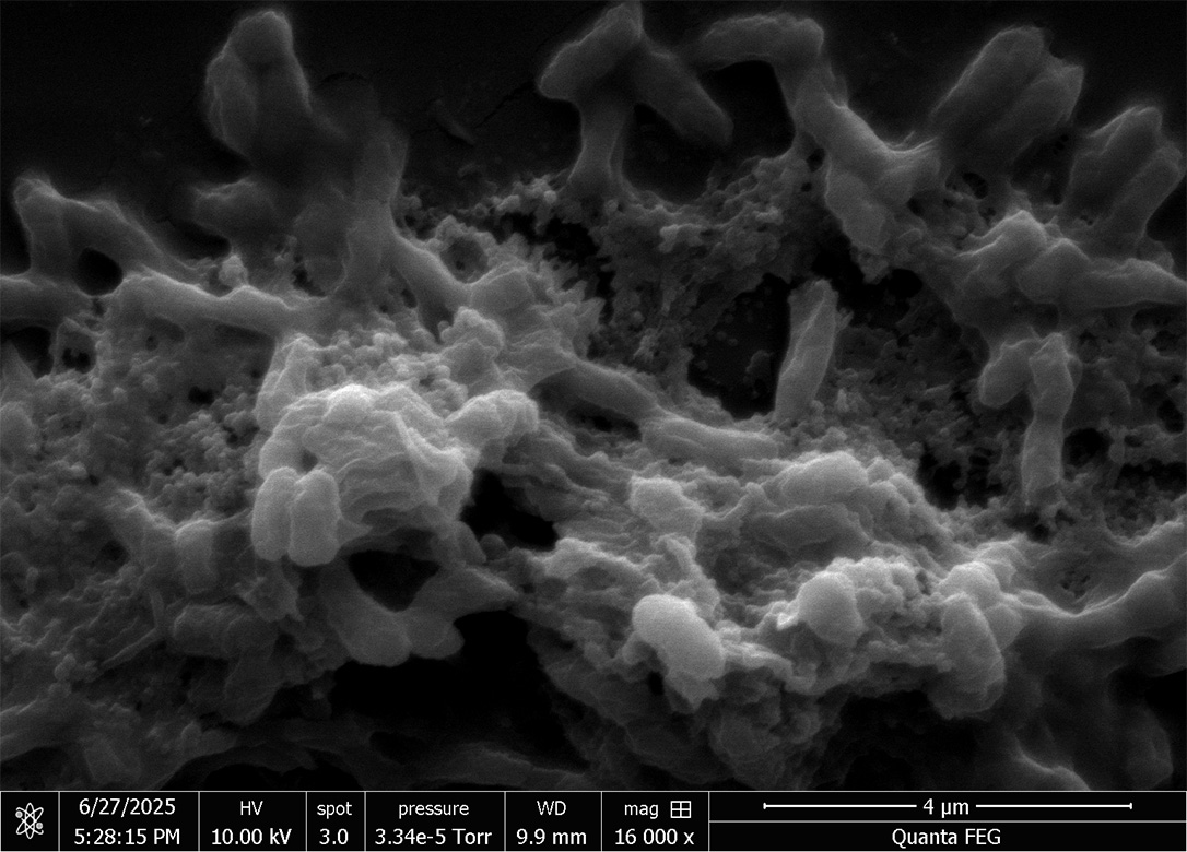
Bacteria are single-celled organisms, but they rarely live alone. Rapidly reproducing and struggling for space in a competitive and sometimes hostile world, they work together to survive. Some bacteria, including Salmonella, form communities embedded in a complex matrix of sugars and proteins that connects and protects them. This biofilm allows the bacteria to share nutrients, transport waste, and resist living and non-living threats in the environment. Biofilms are found in nature, on rocks, soil, and sand. They are also found inside the body, especially on our teeth and in our intestines, and on human-made surfaces.
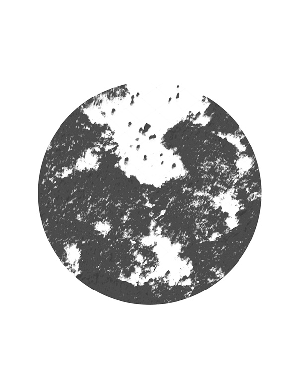
Biofilms of disease-causing Salmonella can be dangerous, in particular because the bacteria forming them are shielded from chemical and physical cleaning techniques. When Salmonella biofilms grow on the synthetic surface of a medical device or food storage container, shed nanoplastics alter the biology of the bacterial cells. Images like this one, in which the bacteria of a biofilm are highlighted in glowing green, allow researchers to track bacterial growth in the presence of plastics and reveal that this exposure can induce them to become more adhesive and more virulent.
SAMFUNDSSIND
As human beings have worked to shape the world around our needs, ants have quietly ruled it. By some estimates, there are more than 20 ant colonies for each living person on Earth. They, like us, make their homes on every continent other than Antarctica; they form societies with sophisticated division of labor, collectively achieving tasks that individuals could not. Among a staggering diversity of species, there are ants that farm aphids or fungus for food, or plants for shelter; ants that teach each other; and ants that manage infectious outbreaks. Sometimes, our societies seek to emulate ants.
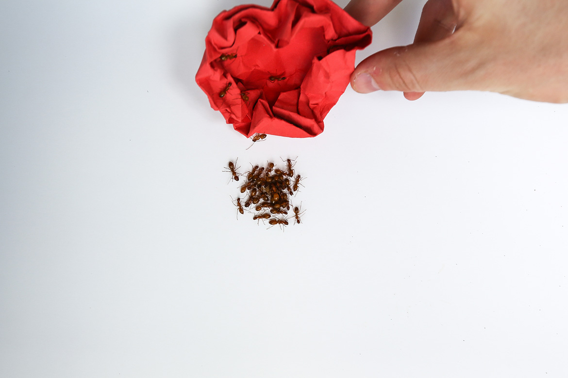
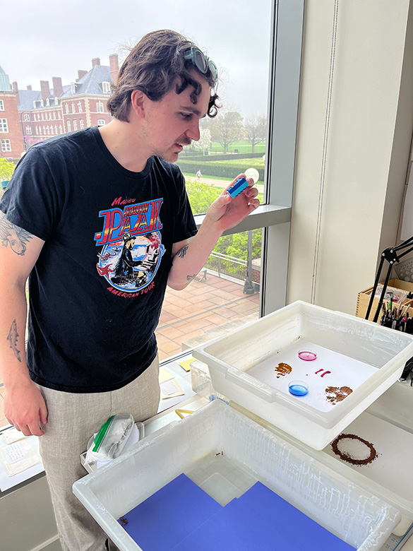
These chestnut carpenter ants live in wooded areas, including those recently disrupted by fire. They have also adapted to our presence; they are one of the most common house-dwelling ants in Illinois, although they do not consume or damage wooden structures. Instead, they make their nests in soft or decaying wood, or in the soil under stones. Shy and nocturnal, the worker ants in these photos are willing to explore novel structures but ultimately seek to re-establish security and order, remaking an echo of their colony structure in safety and out of sight.
SO BUILT THE SCREEN YOU HAD BEGUN
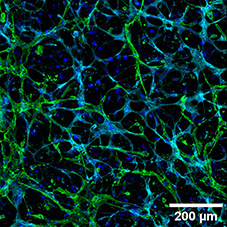
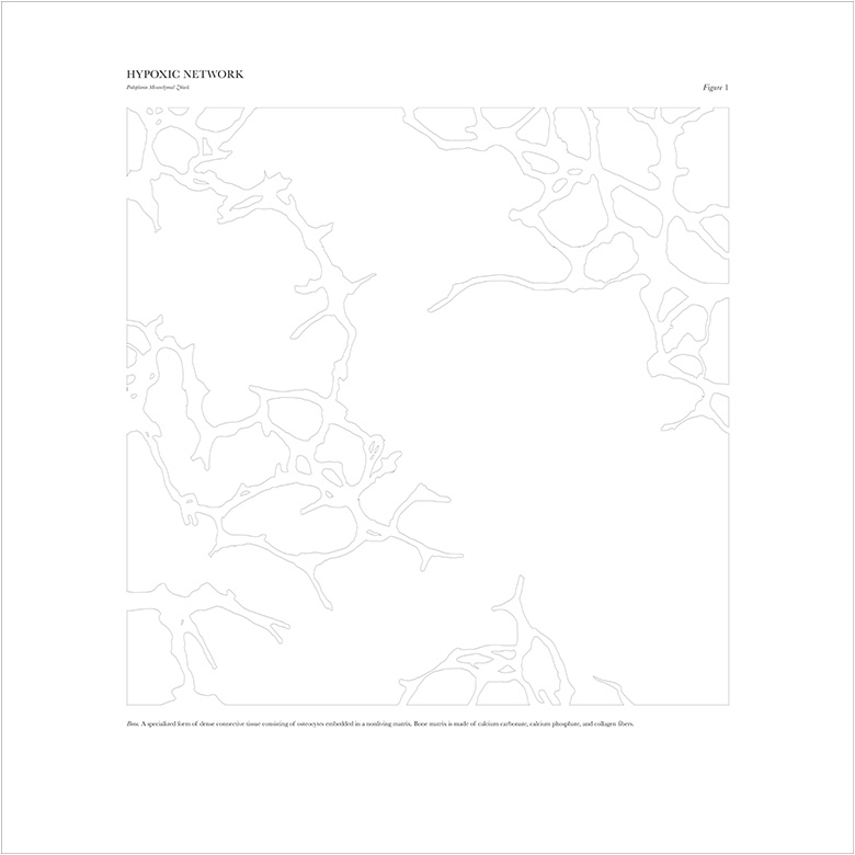
Within the edges of our flat bones are pockets of red marrow, protected nurseries for new blood cells. Stem cells in the marrow divide to replenish the red blood cells, white blood cells, and platelets that circulate throughout our bodies. As these new cells migrate out of the marrow, the support cells that surrounded and fed them remain behind. Together, endothelial cells that line the blood vessels of the marrow and stromal cells beside them provide the stem cells and their daughters with physical support, nutrients, and chemical signals that guide their development.
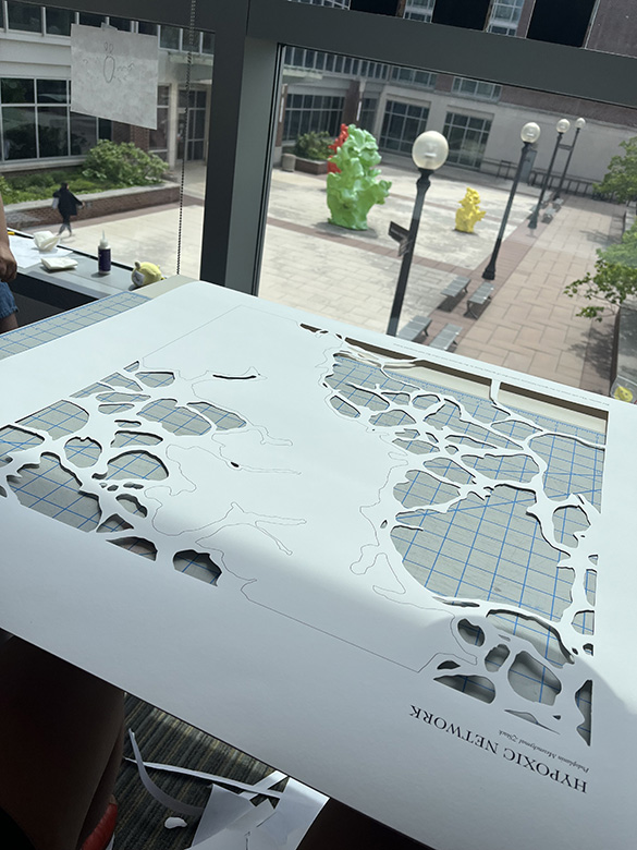
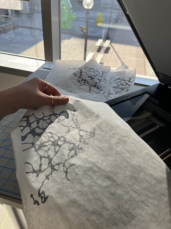
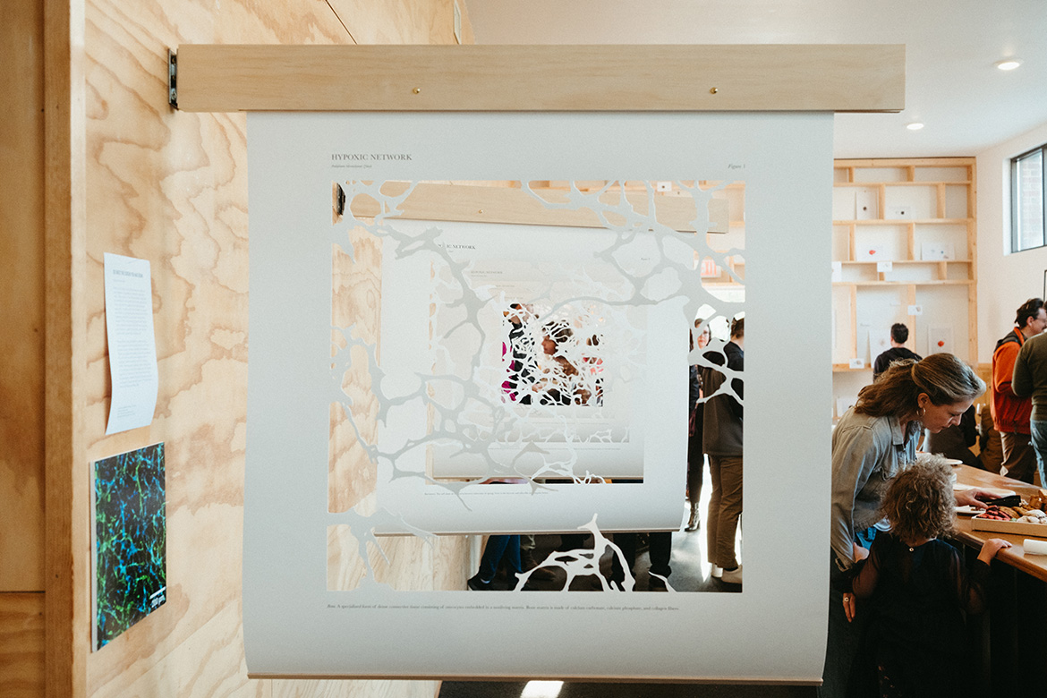
Researchers are interested in understanding these signals, in both healthy marrow and in the disease states of blood disorders like leukemia. These cut-paper silhouettes show the lacy network the cells form as they grow together within a hydrogel matrix designed to mimic a bone’s interior. Microscopic imaging lights up one plane of the cells at a time; the images are then layered together to reconstruct their three-dimensional relationships. Growing the cells this way gives us a window into a process and a part of the body that is vital, but otherwise obscured.
THE FEET OF SHADOWS
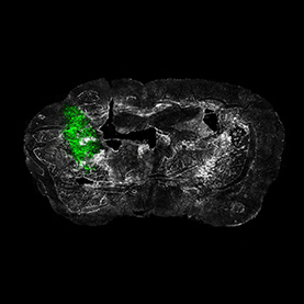
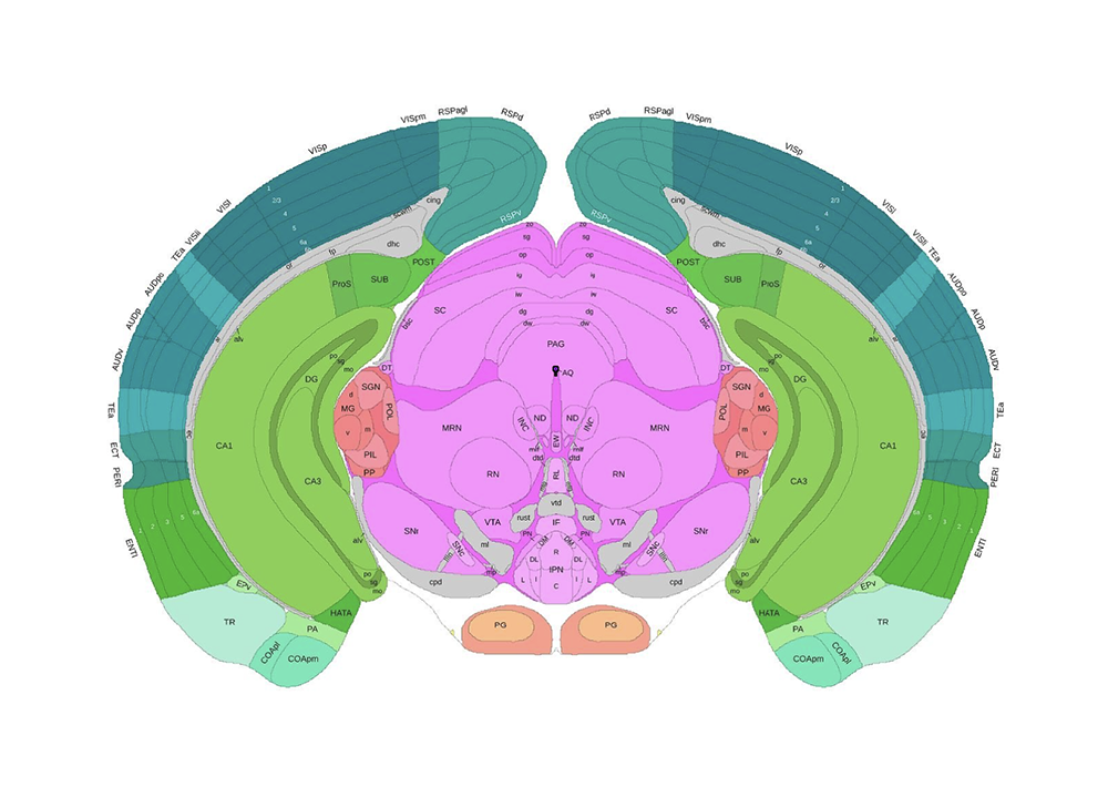
Within the brain, networks of neurons spark with activity, transforming signals from senses to thoughts to actions. Intermingled with these remarkable cells are glia, an equally hardworking but often unrecognized cell type. Glial cells provide support, sustenance, and protection that neurons cannot do without. It is only when something goes wrong that glia receive more attention.
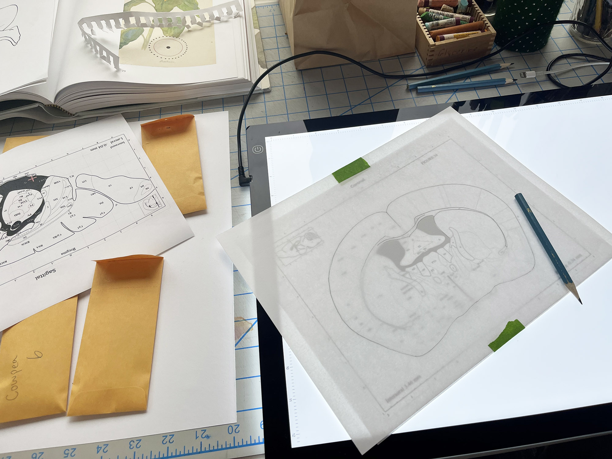
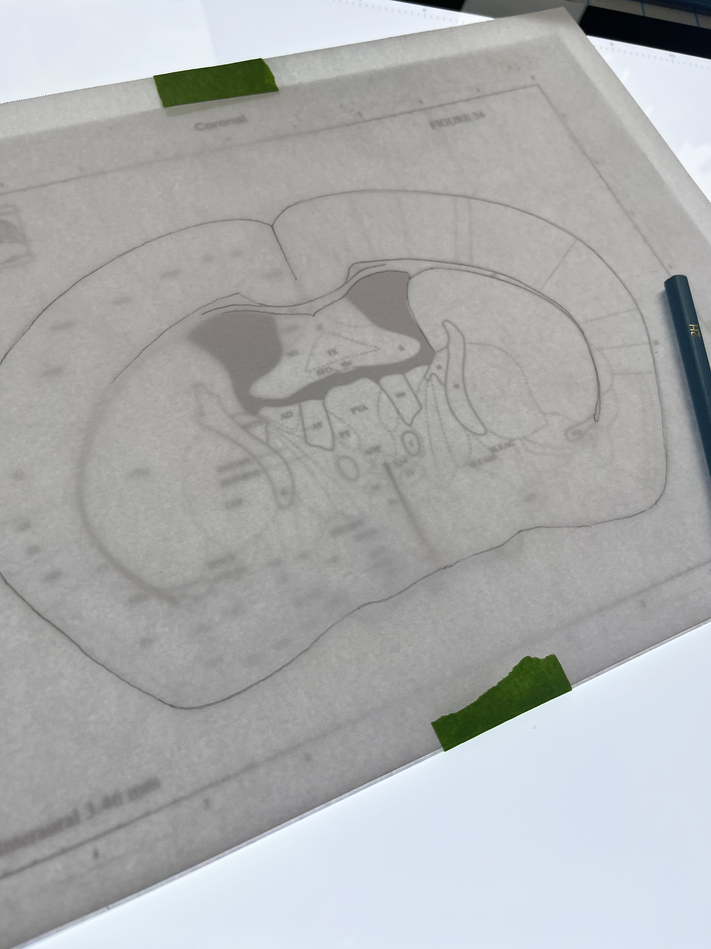
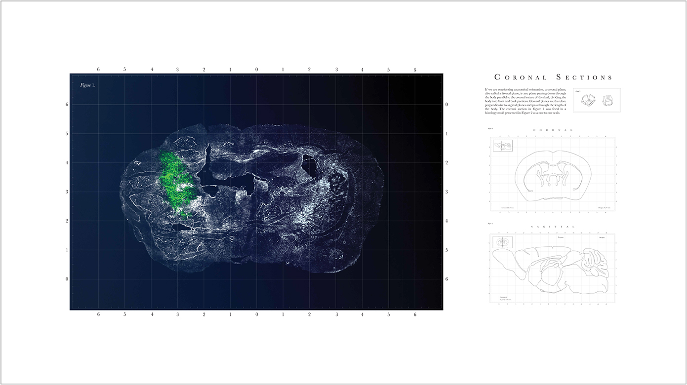
Glioblastoma is a devastatingly aggressive form of brain cancer characterized by the overgrowth of glial cells. Disconnected from their supportive role, the expansion of these cells is insidious as they infiltrate healthy brain tissue; the resulting tumors are almost impossible to completely remove, as they have no clear borders. By examining the anatomy of mouse models of glioblastoma, like the one that yielded the brain section seen here, researchers can learn how to grapple with this deadly disease. A fluorescent dye reveals how the evasive cancer cells invade the healthy tissue surrounding them. As we become able to trace their paths, we may learn to thwart them.
VOLVELLE VENERIS
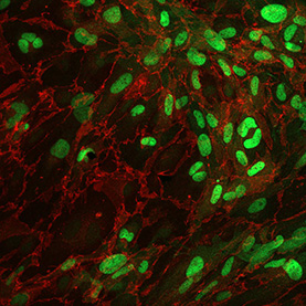
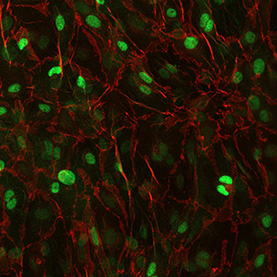
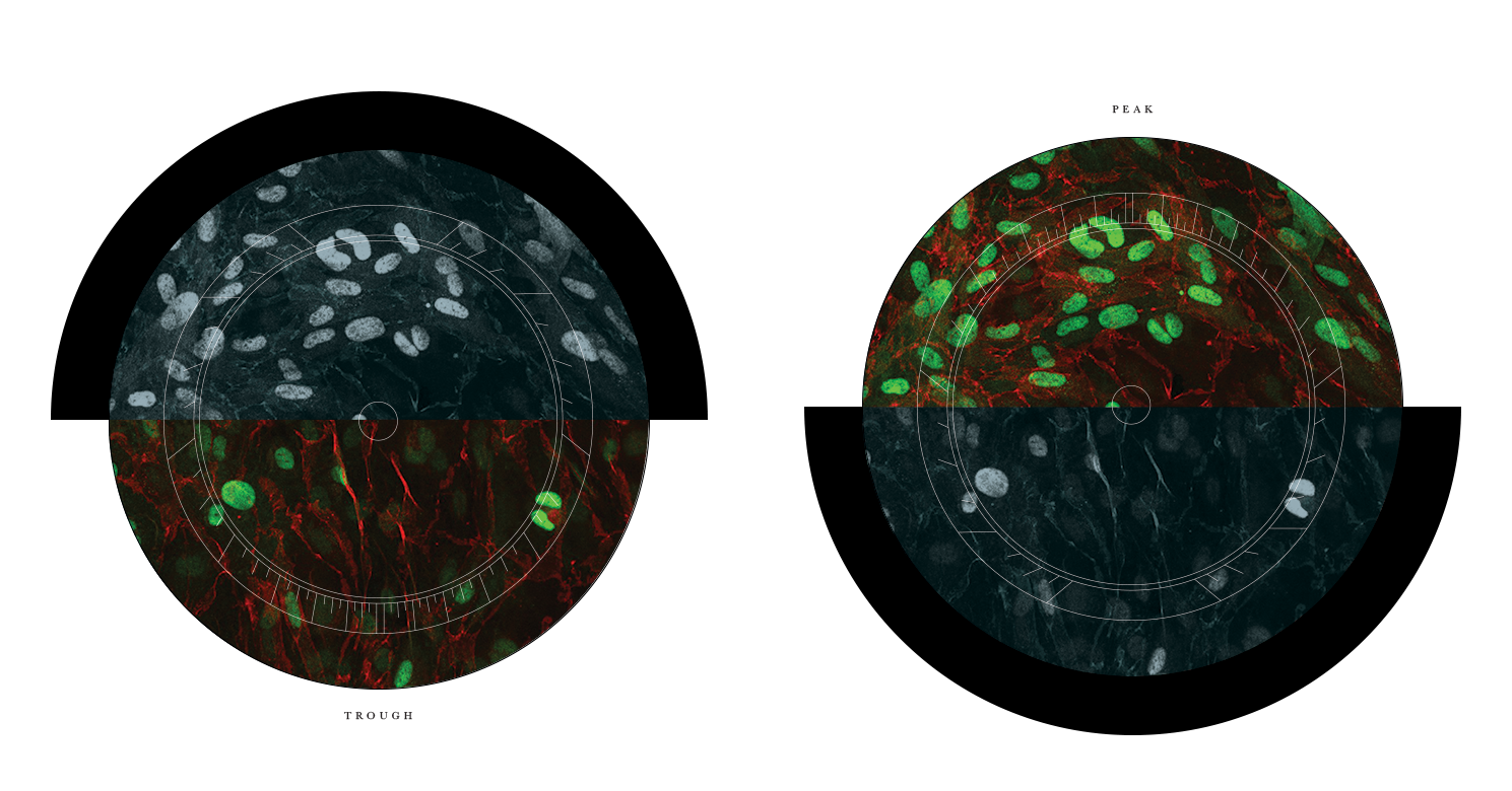
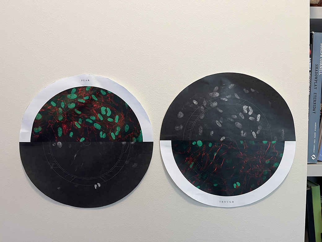
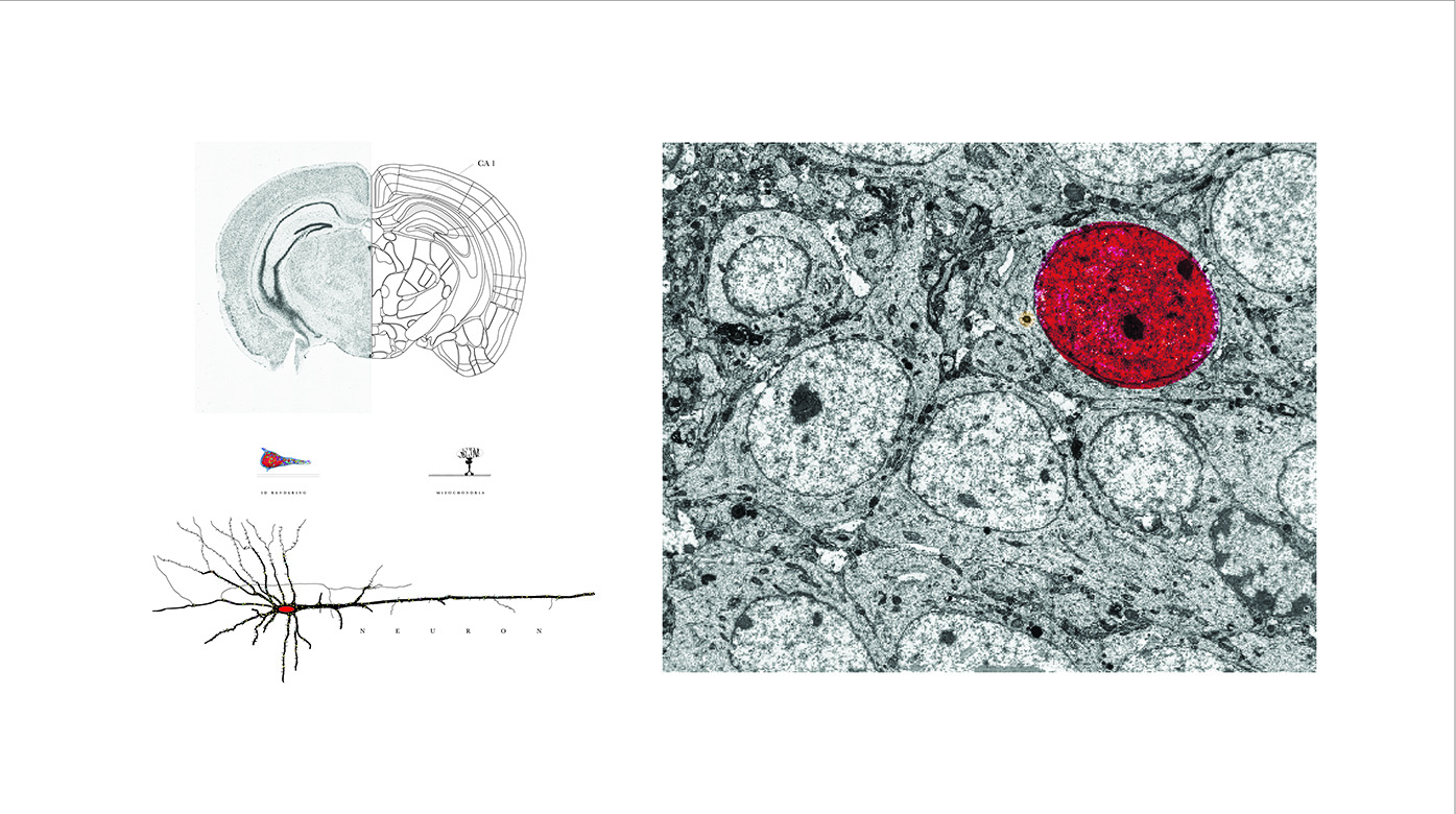
The brain is the central clearinghouse for the vast array of signals our bodies receive from our environment. Regulating the brain’s exposure to the world beyond is a fine balance; it is reliant on nutrients and signals from the body through the bloodstream, but must be protected from toxins and pathogens. The endothelial cells that line the brain’s blood vessels are a key component of this protection. These cells are not like bricks in a wall; they hold their position like a row of people with their arms tightly linked, carefully deciding what may pass through.
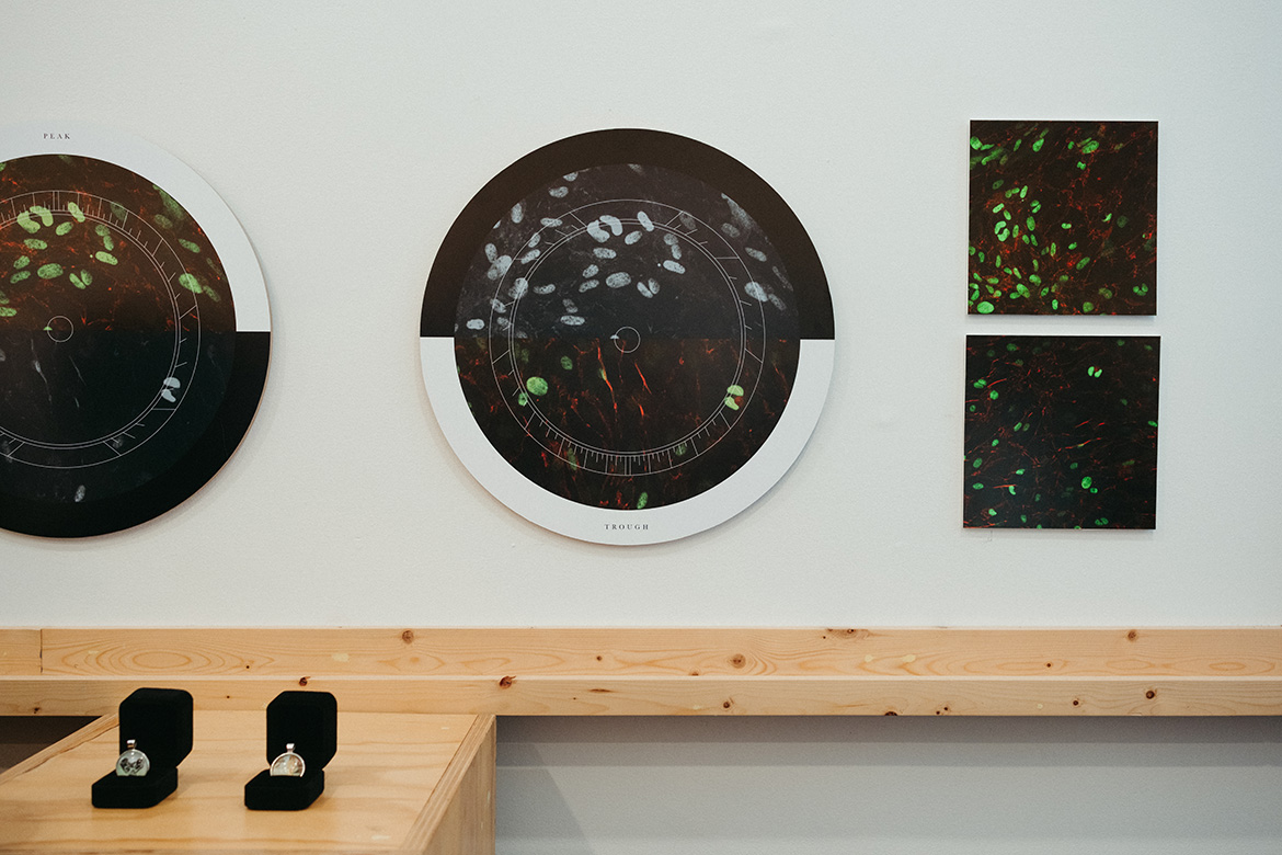
When this barrier is weakened by age or disease, blood can leak into the brain, causing damage and cell death. This type of bleeding is more common in the night and early morning. By studying the blood-brain barrier throughout the circadian cycle, researchers hope to understand this pattern of pathology. These images show endothelial cells producing a glowing green dye called Venus, its intensity reflecting the time on their internal clocks. Grown outside the body for closer study, the cells form clusters, sheets, and—once—the spontaneous silhouette of a microscopic heart.
WALKING THE WARP THREADS
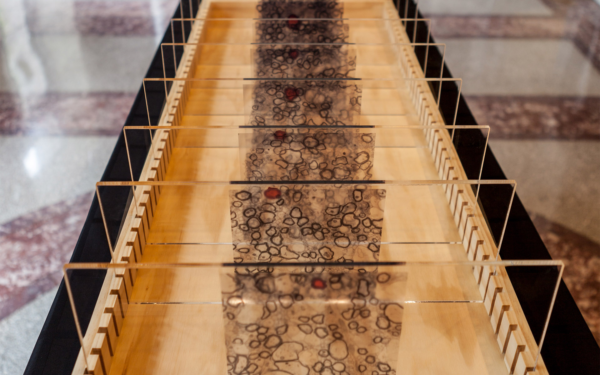
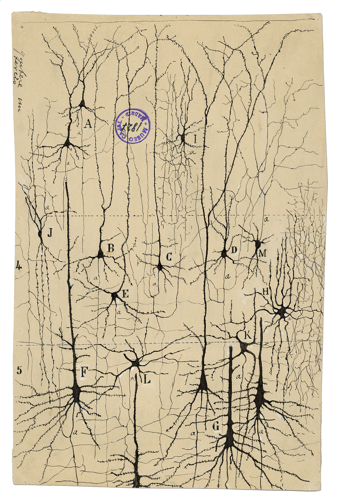
Biological computing power is energetically costly: the human brain makes up just 2% of the human body’s mass, yet it consumes as much as 20% of its calories. Neurons in the nervous system, like all other cells of the body, are supplied with a usable form of chemical energy by tiny internal structures called mitochondria. In the branching structures of a neuron, mitochondria are constantly replenished and shuttled throughout the cell to meet the shifting energy demands of their signaling activity.
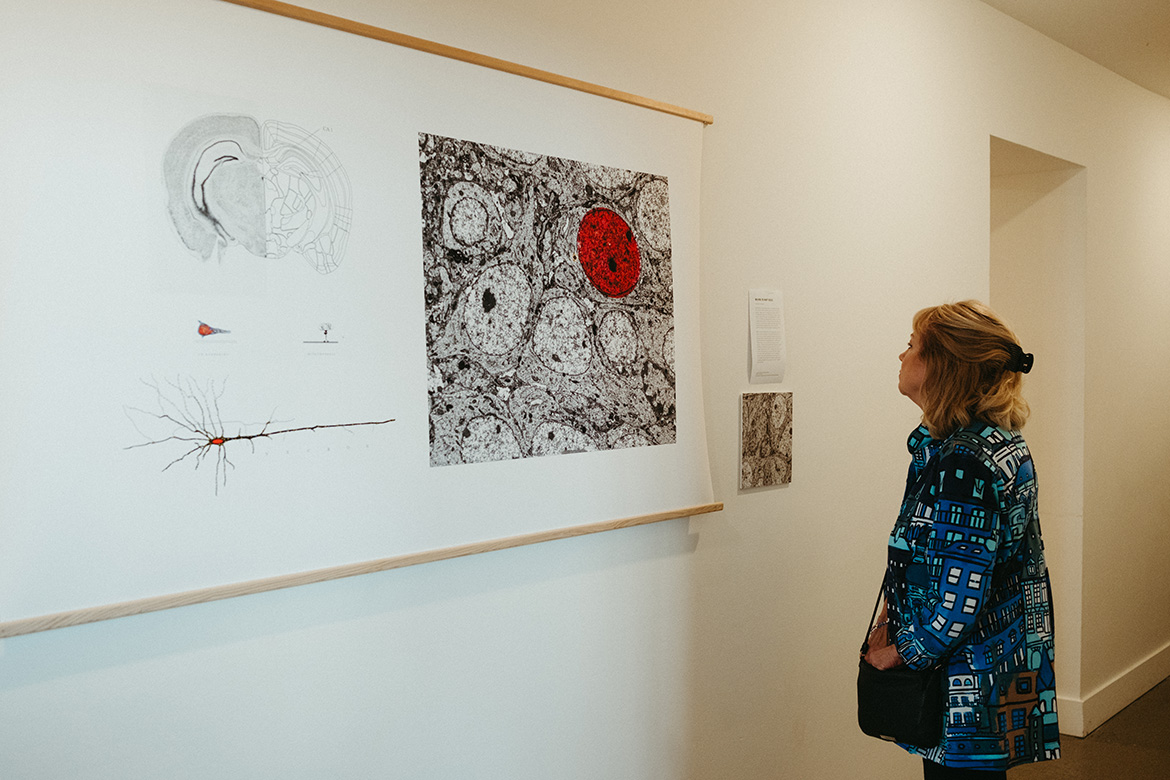
When mitochondria are not working well, neurons lack sufficient power to form new connections or to process and send signals through existing ones. Researchers track the distribution of mitochondria within neurons to understand their function and dysfunction. In a neuron’s microscopic cross-section, they can locate and count the dark, round profiles of individual mitochondria. This set of images relates a neuron’s context within the mouse brain to its isolated structure, with the nucleus in its cell body highlighted in red. Hundreds of serial slices have enabled the reconstruction of the shapes and positions of mitochondria within, revealing details of their dynamic role in health and disease.


View Gallery
1
/
10
1 / x